Bone and Soft Tissue for NEET PG Pathology: Osteomyelitis, Bone Tumours, Paget Disease, Rheumatoid Arthritis, Osteoarthritis, Gout, Sarcoma
Start Here
🎯 Exam priority: Medium–high. Expect: bone tumour by age and site (epiphysis vs metaphysis), Codman triangle and onion-skin periosteum, Ewing translocation, osteomyelitis organisms and sites, Paget disease markers and sarcoma risk, RA vs OA joints, gout vs CPPD crystals, and soft tissue sarcoma translocations.
CBME mapping: PA 32.1–32.7. Rickets and osteomalacia are in article 8; myeloma in article 13; translocations in article 4.
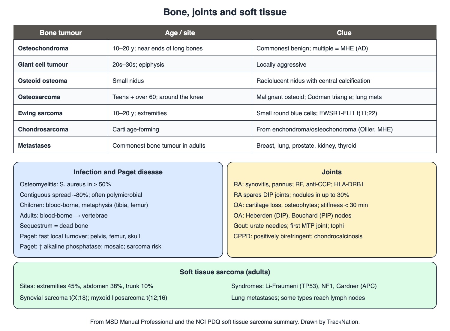
Bone tumours, infection, Paget disease, joints and soft tissue sarcoma. Drawn by TrackNation.
Osteomyelitis and Paget Disease
Osteomyelitis: about 80% from contiguous spread (wounds, nearby infection), often polymicrobial; the rest blood-borne or from open fractures and surgery. S. aureus is present in at least half. Blood-borne disease in children hits the metaphyses of tibia, femur or humerus; in adults it hits the vertebrae. Adult risks: age, haemodialysis, sickle cell disease, catheters, immunosuppression, injection drug use. Dead bone = sequestrum.
Paget disease of bone: late-onset, localised, accelerated bone turnover (overactive osteoclasts, NF-κB; SQSTM1 variants) replacing normal matrix with soft, enlarged bone. Pelvis, femur and skull most often. Raised alkaline phosphatase; skull X-ray shows a mosaic of sclerosis and lucency. New or severe pain suggests osteosarcoma.
Bone Tumours
Metastases far outnumber primary malignant bone tumours in adults — from breast, lung, prostate, kidney, thyroid and colon.
Osteochondroma (exostosis): the commonest benign bone tumour; ages 10–20; near the ends of long bones; multiple ones = multiple hereditary exostoses (autosomal dominant).
Giant cell tumour: 20s–30s; epiphyseal (and distal metaphyseal-epiphyseal); locally aggressive. Osteoid osteoma: radiolucent nidus with central calcification. Aneurysmal bone cyst: expansile, usually before 25.
Osteosarcoma: the commonest primary malignant bone tumour (counting myeloma as marrow). Peaks in adolescence (growth spurt) and after 60 (Paget, bone infarcts, prior radiation). Malignant osteoid; around the knee (distal femur > proximal tibia), metaphyseal-diaphyseal; Codman triangle; spreads to lung or other bone.
Ewing sarcoma: small round blue cell tumour, peak 10–20 years, mostly extremities; layered periosteal new bone gives an onion-skin look; EWSR1-FLI1 fusion from t(11;22) in about 85%.
Chondrosarcoma: malignant cartilage-forming tumour; mostly primary, sometimes arising from an enchondroma or osteochondroma (Ollier disease, Maffucci syndrome, multiple hereditary exostoses).
Joints
Rheumatoid arthritis: autoimmune synovitis; genetic link to the HLA-DRB1 shared epitope; smoking may contribute. Hyperplastic synovium (pannus) erodes cartilage and bone. RF and anti-CCP antibodies. Spares DIP joints. Rheumatoid nodules (granulomas) in up to 30%.
Osteoarthritis: progressive cartilage loss with osteophytes and sclerosis; pain on use; morning stiffness under 30 minutes; non-inflammatory fluid. Heberden (DIP) and Bouchard (PIP) nodes.
Gout: monosodium urate crystals; first attack usually one joint, often the first MTP; urate > 6.8 mg/dL; needle-shaped birefringent crystals; tophi with a granulomatous rim. CPPD: positively birefringent crystals; chondrocalcinosis, commoner with age.
Feature | Rheumatoid arthritis | Osteoarthritis | Gout |
Core lesion | Synovitis with pannus | Cartilage loss, osteophytes | Urate crystal deposition |
Joints | Any except DIP | DIP (Heberden), PIP (Bouchard), thumb base | First MTP joint first |
Lab / fluid | RF, anti-CCP | Non-inflammatory (≤ 2000 WBC/µL) | Needle-shaped birefringent crystals |
Extra | Nodules in up to 30%; HLA-DRB1 | Morning stiffness < 30 min | Tophi with granulomatous rim |
Soft Tissue Sarcoma
Adult sites: extremities 45%, intra-abdominal 38%, trunk 10%, head and neck 5%.
Syndromes: Li-Fraumeni (TP53), neurofibromatosis 1 (NF1), Gardner (APC).
Signature translocations: synovial sarcoma t(X;18); myxoid liposarcoma t(12;16). Undifferentiated pleomorphic sarcoma was formerly called malignant fibrous histiocytoma. Synovial, clear cell, angiosarcoma, rhabdomyosarcoma and epithelioid sarcoma tend to reach lymph nodes.
Traps the Examiner Sets
Epiphysis in a 20–30-year-old = giant cell tumour; metaphysis around the knee in a teenager = osteosarcoma.
Onion skin = Ewing; Codman triangle = osteosarcoma.
Child osteomyelitis = metaphysis; adult blood-borne = vertebra.
RA spares DIP; OA loves DIP (Heberden).
Gout = needles; CPPD = positively birefringent.
Memory Hooks & Shortcuts
Shortcuts to lock in this article's numbers and lists. Each one only re-packages facts proven above.
🧠 Giant cell tumour = Grown-ups' Epiphysis (20–30s) — GCT
🧠 Ewing = Eleven-Twenty-two, onion skin — Ewing sarcoma
🧠 Osteosarcoma = Osteoid + Codman + Knee — osteosarcoma
🧠 Paget = Pelvis, Femur, Skull + ALP — Paget disease
🧠 Gout Needles, CPPD Positive — crystals
🧠 Synovial = X and 18 (Xtra-eighteen) — synovial sarcoma
Rapid-Revision Cards
Osteomyelitis organism
S. aureus ≥ 50%.
Osteomyelitis route
Contiguous ~80%; blood-borne rest.
Child vs adult site
Child metaphysis; adult vertebra.
Sequestrum
Dead bone.
Osteochondroma
Commonest benign; 10–20 y; MHE.
Giant cell tumour
20s–30s; epiphysis; locally aggressive.
Osteoid osteoma
Radiolucent nidus, central calcification.
Osteosarcoma
Teens + > 60; knee; osteoid; Codman; lung mets.
Ewing
10–20 y; small round blue cells; onion skin; t(11;22).
Chondrosarcoma
Cartilage; from enchondroma/osteochondroma.
Bone metastases
Commonest in adults; breast, lung, prostate.
Paget of bone
Osteoclasts; ALP ↑; mosaic; sarcoma risk.
RA
Pannus; RF, anti-CCP; HLA-DRB1; spares DIP.
RA nodules
Up to 30%.
OA
Cartilage loss; osteophytes; Heberden/Bouchard.
Gout
Needle urate crystals; first MTP; tophi.
CPPD
Positively birefringent; chondrocalcinosis.
Synovial sarcoma
t(X;18).
Myxoid liposarcoma
t(12;16).
Sarcoma syndromes
Li-Fraumeni, NF1, Gardner.
Sources
Facts in this article were checked against these documents.
MSD Manual Professional: Osteomyelitis; Benign Bone Tumors and Cysts; Primary Malignant Bone Tumors; Metastatic Bone Tumors; Paget Disease of Bone — Merck & Co. (MSD Manuals), reviewed 2025–26 (link)
MSD Manual Professional: Rheumatoid Arthritis; Osteoarthritis; Gout; Calcium Pyrophosphate Arthritis — Merck & Co. (MSD Manuals), reviewed 2025–26 (link)
PDQ Adult Soft Tissue Sarcoma Treatment (Health Professional Version) — US National Cancer Institute, 2026 (link)
Competency Based Undergraduate Curriculum, Pathology (PA 32) — National Medical Commission, 2024
Test Yourself: Practice Questions
Thirty questions in NEET PG style, most of them clinical vignettes.
Practice Questions (NEET PG pattern)
Original questions written in the style of the exam: +4 for a correct answer, −1 for a wrong one. Every answer was checked against the sources listed above. NEET PG papers are not published by NBEMS, so these are not past-paper questions.
Q1. The organism present in at least half of osteomyelitis cases is
(a) Staphylococcus aureus
(b) Pseudomonas
(c) Salmonella
(d) Mycobacterium tuberculosis
Show answer
Answer: (a) — Including MRSA.
Q2. In a child, blood-borne osteomyelitis most often affects the
(a) skull
(b) epiphysis only
(c) vertebral body
(d) metaphysis of a long bone such as the tibia or femur
Show answer
Answer: (d) — In adults, blood-borne infection usually hits the vertebrae.
Q3. A haemodialysis patient develops blood-borne osteomyelitis. The usual site in adults is the
(a) distal femoral metaphysis
(b) phalanges
(c) vertebrae
(d) skull
Show answer
Answer: (c) — Haemodialysis is a listed adult risk factor.
Q4. A fragment of dead bone within an area of chronic osteomyelitis is called
(a) a sequestrum
(b) a Brodie abscess
(c) a tophus
(d) an involucrum
Show answer
Answer: (a) — Non-viable bone.
Q5. Most osteomyelitis arises by
(a) blood-borne spread (~80%)
(b) lymphatic spread
(c) contiguous spread from nearby infected tissue or wounds (~80%)
(d) spread along nerves
Show answer
Answer: (c) — Often polymicrobial.
Q6. A 15-year-old has a lump near the end of a long bone; X-ray shows an osteocartilaginous exostosis. The lesion and its status are
(a) osteosarcoma — malignant
(b) osteochondroma — the commonest benign bone tumour
(c) giant cell tumour — epiphyseal
(d) Ewing sarcoma
Show answer
Answer: (b) — Multiple ones = multiple hereditary exostoses.
Q7. A 28-year-old has a locally aggressive lesion in the epiphysis of a long bone. The likely tumour is
(a) osteosarcoma
(b) giant cell tumour of bone
(c) Ewing sarcoma
(d) osteochondroma
Show answer
Answer: (b) — Peak in the 20s and 30s.
Q8. CT of a painful small bone lesion shows a radiolucent nidus with central calcification. The diagnosis is
(a) enchondroma
(b) osteochondroma
(c) aneurysmal bone cyst
(d) osteoid osteoma
Show answer
Answer: (d) — Treated by radiofrequency ablation or curettage.
Q9. Multiple osteochondromas inherited in an autosomal dominant pattern are called
(a) multiple hereditary exostoses
(b) Maffucci syndrome
(c) Ollier disease
(d) fibrous dysplasia
Show answer
Answer: (a) — Ollier disease is multiple enchondromas.
Q10. A 16-year-old has pain and swelling above the knee; X-ray shows a destructive distal femoral lesion with a Codman triangle. Biopsy will show
(a) small round blue cells with t(11;22)
(b) malignant cells producing osteoid
(c) giant cells in an epiphyseal lesion
(d) cartilage only
Show answer
Answer: (b) — Osteosarcoma; spreads to lung or other bone.
Q11. The second age peak of osteosarcoma, after 60, is linked to
(a) gout
(b) multiple hereditary exostoses only
(c) Paget disease, bone infarcts and prior radiation
(d) rickets
Show answer
Answer: (c) — The first peak matches the adolescent growth spurt.
Q12. Osteosarcoma most commonly metastasises to the
(a) liver
(b) lungs (and other bones)
(c) lymph nodes first
(d) brain
Show answer
Answer: (b) — Chest CT is part of staging.
Q13. A 13-year-old has a long-bone tumour with layered 'onion-skin' periosteal reaction; biopsy shows small round blue cells. The typical genetic change is
(a) t(11;22) producing EWSR1-FLI1
(b) t(12;16)
(c) t(X;18)
(d) BRAF V600E
Show answer
Answer: (a) — Ewing sarcoma; the fusion is present in about 85%.
Q14. The peak age of Ewing sarcoma is
(a) 10–20 years
(b) 40–60 years
(c) under 2 years
(d) over 70 years
Show answer
Answer: (a) — Mostly in the extremities.
Q15. A malignant cartilage-forming bone tumour that may arise from a pre-existing enchondroma is
(a) osteosarcoma
(b) chordoma
(c) Ewing sarcoma
(d) chondrosarcoma
Show answer
Answer: (d) — Especially in Ollier disease, Maffucci syndrome or MHE.
Q16. In adults, the commonest malignant tumour found in bone is
(a) Ewing sarcoma
(b) osteosarcoma
(c) metastatic carcinoma
(d) chondrosarcoma
Show answer
Answer: (c) — Breast, lung, prostate, kidney, thyroid, colon.
Q17. An older man has an enlarging skull and bone pain; alkaline phosphatase is high and skull X-ray shows a mosaic of sclerosis and lucency. The disease is
(a) Paget disease of bone
(b) multiple myeloma
(c) osteoporosis
(d) osteomalacia
Show answer
Answer: (a) — Accelerated local bone turnover; pelvis, femur, skull.
Q18. In Paget disease of bone, new severe pain at a pagetic site should raise suspicion of
(a) osteoid osteoma
(b) gout
(c) septic arthritis
(d) osteosarcoma
Show answer
Answer: (d) — A rare but serious complication.
Q19. The cell whose overactivity drives Paget disease of bone is the
(a) chondrocyte
(b) osteocyte
(c) plasma cell
(d) osteoclast
Show answer
Answer: (d) — Increased NF-κB activity; SQSTM1 variants.
Q20. A woman has symmetrical small-joint polyarthritis sparing the distal interphalangeal joints; anti-CCP is positive. The destructive tissue in her joints is
(a) osteophytes
(b) urate tophi
(c) hyperplastic synovium (pannus)
(d) calcium pyrophosphate deposits
Show answer
Answer: (c) — Rheumatoid arthritis.
Q21. The genetic predisposition to rheumatoid arthritis maps to
(a) a shared epitope in HLA-DRB1
(b) HLA-A3
(c) HLA-B27
(d) HLA-DQ2
Show answer
Answer: (a) — Class II histocompatibility antigens.
Q22. Subcutaneous nodules occur in up to what share of patients with rheumatoid arthritis?
(a) 1%
(b) never
(c) 90%
(d) 30%
Show answer
Answer: (d) — They are granulomas; prevalence is declining.
Q23. An older woman has hard bony swellings of the distal interphalangeal joints and pain after use with brief morning stiffness. The nodes are
(a) Heberden nodes of osteoarthritis
(b) rheumatoid nodules
(c) tophi
(d) Bouchard nodes of RA
Show answer
Answer: (a) — Bouchard nodes are at the PIP joints.
Q24. The core pathology of osteoarthritis is
(a) progressive loss of joint cartilage with osteophytes and sclerosis
(b) bacterial infection
(c) autoimmune synovitis with pannus
(d) crystal deposition
Show answer
Answer: (a) — Synovial fluid is non-inflammatory.
Q25. A man has an acutely painful, red, swollen first metatarsophalangeal joint. Joint fluid would show
(a) positively birefringent rhomboid crystals
(b) gram-positive cocci always
(c) needle-shaped birefringent urate crystals
(d) cholesterol crystals
Show answer
Answer: (c) — Gout; the first attack is usually one joint.
Q26. Chalky deposits of urate crystals surrounded by a granulomatous reaction in gout are
(a) tophi
(b) rheumatoid nodules
(c) Heberden nodes
(d) sequestra
Show answer
Answer: (a) — Most often in joints and skin.
Q27. An elderly patient has calcification of the knee cartilage and positively birefringent crystals in joint fluid. The disorder is
(a) rheumatoid arthritis
(b) calcium pyrophosphate deposition (chondrocalcinosis)
(c) septic arthritis
(d) gout
Show answer
Answer: (b) — Commoner with age.
Q28. A soft tissue sarcoma with translocation t(X;18) is
(a) synovial sarcoma
(b) Ewing sarcoma
(c) rhabdomyosarcoma
(d) myxoid liposarcoma
Show answer
Answer: (a) — Myxoid liposarcoma has t(12;16).
Q29. In adults, soft tissue sarcomas most often arise in the
(a) heart
(b) head and neck
(c) extremities (45%)
(d) trunk only
Show answer
Answer: (c) — Then intra-abdominal organs (38%).
Q30. An inherited syndrome predisposing to soft tissue sarcoma is
(a) Turner syndrome
(b) Li-Fraumeni syndrome (TP53)
(c) Marfan syndrome
(d) Down syndrome
Show answer
Answer: (b) — Also NF1 and Gardner syndrome.
Active Recall
From memory: describe osteomyelitis routes, organisms and sites by age; place each bone tumour by age and site and give its hallmark; explain Paget disease; contrast RA, OA, gout and CPPD; and give soft tissue sarcoma sites, syndromes and translocations.