Human Reproduction — Part A (NEET Biology Class 12): Reproductive Systems & Gametogenesis
The Male Reproductive System
🎯 NEET priority: High-yield. Part A of Human Reproduction — the reproductive systems and gametogenesis. Gametogenesis diagrams (spermatogenesis vs oogenesis) are a NEET perennial — expect a question almost every year.
Humans are sexually reproducing and viviparous. The full reproductive sequence: gametogenesis (sperm/ovum formation) → insemination (sperm transfer) → fertilisation (gamete fusion → zygote) → implantation → gestation → parturition. Notably, sperm formation continues into old age in men, but ovum formation ceases around age 50 in women.
The male reproductive system
The testes sit in the scrotum, outside the abdomen, at a temperature 2–2.5°C lower than body temperature — essential for spermatogenesis. Each testis (4–5 cm × 2–3 cm) has about 250 testicular lobules, each with 1–3 highly coiled seminiferous tubules — the site of sperm production.
The seminiferous tubule wall has two cell types: spermatogonia (germ cells, undergo meiosis to form sperm) and Sertoli cells (provide nutrition to germ cells — do NOT make sperm themselves). Outside the tubules, Leydig (interstitial) cells synthesise androgens (testicular hormones).
Sperm path: seminiferous tubule → rete testis → vasa efferentia → epididymis → vas deferens → ejaculatory duct → urethra. Accessory glands — paired seminal vesicles, a single prostate, paired bulbourethral glands — secrete the fructose- and calcium-rich seminal plasma.
The Female Reproductive System & Spermatogenesis
The female reproductive system
Ovaries (2–4 cm) produce the ovum and steroid hormones. Female accessory ducts: paired fallopian tubes/oviducts (10–12 cm — infundibulum with fimbriae → ampulla, the site of fertilisation → isthmus), a single uterus, and the vagina.
The uterine wall has 3 layers: outer perimetrium, middle muscular myometrium (contracts strongly at delivery), inner glandular endometrium (undergoes cyclical change during menstruation). Mammary glands have 15–20 mammary lobes of milk-secreting alveoli.
Trap: presence or absence of the hymen is NOT a reliable indicator of virginity or sexual experience — it can tear from sport, a fall, or a tampon, and sometimes persists after coitus.
Spermatogenesis
A spermatogonium is diploid (46 chromosomes) and multiplies by mitosis. A primary spermatocyte undergoes meiosis I → two haploid secondary spermatocytes (23 chromosomes each) → meiosis II → four haploid spermatids. Spermatids transform into sperm by spermiogenesis; release from the tubule is spermiation.
Hormonal control: GnRH (hypothalamus) → LH + FSH (anterior pituitary). LH acts on Leydig cells → androgen secretion → drives spermatogenesis. FSH acts on Sertoli cells → factors that aid spermiogenesis.
Sperm structure: head (haploid nucleus + acrosome cap of enzymes for fertilisation), neck, middle piece (packed with mitochondria — energy for tail movement), tail. One ejaculation releases 200–300 million sperms; normal fertility needs ≥60% normal shape/size and ≥40% vigorous motility.
Oogenesis & Why This Matters for NEET
Oogenesis
Oogenesis starts in the fetal ovary — millions of oogonia form, and no new oogonia form after birth. They enter meiosis I and arrest as primary oocytes, each surrounded by granulosa cells to form a primary follicle. Most degenerate; only 60,000–80,000 primary follicles remain per ovary at puberty.
A primary follicle → secondary follicle → tertiary follicle (develops a fluid-filled antrum) → Graafian (mature) follicle. The primary oocyte completes meiosis I inside the tertiary follicle, an unequal division giving a large haploid secondary oocyte + a tiny first polar body. The secondary oocyte develops the zona pellucida and is released at ovulation when the Graafian follicle ruptures.
Spermatogenesis vs oogenesis — side by side
Feature | Spermatogenesis | Oogenesis |
Starts | At puberty | During fetal (embryonic) development |
Continues | Lifelong, even in old age | Stops around age 50 (menopause) |
Division of cytoplasm | Equal — 4 equal spermatids per primary spermatocyte | Unequal — 1 large secondary oocyte + tiny polar bodies |
Product count per parent cell | 4 functional sperm | 1 functional ovum (+ polar bodies, which degenerate) |
Meiosis I completed | Continuously, from puberty onward | Only at/near ovulation, decades after being arrested |
Hormonal control of gametogenesis — who acts on what
Hormone | Source | Target | Effect |
GnRH | HYPOTHALAMUS | Anterior pituitary | Triggers the whole cascade at puberty — stimulates LH and FSH secretion |
LH | Anterior pituitary | LEYDIG cells (male) | Stimulates synthesis and secretion of ANDROGENS, which drive spermatogenesis |
FSH | Anterior pituitary | SERTOLI cells (male) | Stimulates secretion of factors that help SPERMIOGENESIS |
Androgens | Leydig cells | Seminiferous tubules and accessory ducts/glands | Drive spermatogenesis; maintain accessory duct and gland function |
Male vs female reproductive system — side by side
Feature | Male | Female |
Primary sex organ | Testis — in the SCROTUM, outside the abdomen (needs 2–2.5°C cooler) | Ovary — inside the lower abdomen |
Gamete production site | Seminiferous tubules | Ovarian follicles in the cortex |
Nourishing cells | SERTOLI cells (inside the tubule) | GRANULOSA cells (surrounding the oocyte) |
Hormone-secreting cells | LEYDIG/interstitial cells (outside the tubule) → androgens | Follicle and corpus luteum → estrogen and progesterone |
Accessory ducts | Rete testis → vasa efferentia → epididymis → vas deferens → urethra | Fallopian tube (infundibulum → ampulla → isthmus) → uterus → vagina |
Gametes produced per parent cell | 4 functional sperm | 1 functional ovum (+ polar bodies) |
Timing | Starts at puberty, continues into old age | Oogonia fixed BEFORE birth; ceases around age 50 |
Why this matters for NEET
High-value one-liners: Sertoli cells nourish germ cells, they do NOT secrete androgens (Leydig cells do — a classic True/False NCERT exercise trap); spermatogonium (46) → secondary spermatocyte (23, after meiosis I) → spermatid (23, after meiosis II, but 4 formed per primary spermatocyte); oogenesis is unequal division (secondary oocyte keeps the cytoplasm, polar bodies don't); oogonia are fixed for life — set before birth, unlike sperm made continuously.
Trap: oogenesis's first meiotic division happens inside the tertiary follicle, at/near puberty — NOT completed at the fetal stage (only initiated then, then arrested in prophase I for years/decades). Don't confuse "oogenesis starts in the fetus" with "oogenesis finishes in the fetus."
Test Yourself: MCQs, PYQs & Active Recall
Answer these, then close the article and do an Active Recall. Reveal each answer only after you commit to one.
Practice Questions
Q1. The scrotum maintains testicular temperature at:
(a) The same as body temperature
(b) 10°C lower than body temperature
(c) 2–2.5°C lower than body temperature
(d) 5°C higher than body temperature
Show answer
Answer: (c) — The scrotum keeps testes 2–2.5°C cooler than internal body temperature, essential for spermatogenesis.
Q2. Inside a seminiferous tubule, nutrition to the dividing germ cells is provided by:
(a) Spermatogonia
(b) Sertoli cells
(c) Leydig cells
(d) Epididymal cells
Show answer
Answer: (b) — Sertoli cells line the seminiferous tubule and nourish the germ cells; they do not secrete androgens.
Q3. Androgens are synthesised and secreted by:
(a) Spermatogonia
(b) Sertoli cells
(c) Prostate gland
(d) Leydig (interstitial) cells
Show answer
Answer: (d) — Leydig or interstitial cells, located outside the seminiferous tubules, synthesise testicular androgens.
Q4. The correct path of sperm transport from the testis is:
(a) Vasa efferentia → rete testis → epididymis → vas deferens
(b) Vas deferens → epididymis → rete testis → vasa efferentia
(c) Epididymis → rete testis → vasa efferentia → vas deferens
(d) Rete testis → vasa efferentia → epididymis → vas deferens
Show answer
Answer: (d) — Sperm travel: seminiferous tubule → rete testis → vasa efferentia → epididymis → vas deferens.
Q5. The site of fertilisation within the fallopian tube is the:
(a) Infundibulum
(b) Ampulla
(c) Fimbriae
(d) Isthmus
Show answer
Answer: (b) — The ampulla, the wider part of the oviduct, is where fertilisation normally occurs.
Q6. The three layers of the uterine wall, from outside in, are:
(a) Endometrium, myometrium, perimetrium
(b) Perimetrium, endometrium, myometrium
(c) Myometrium, perimetrium, endometrium
(d) Perimetrium, myometrium, endometrium
Show answer
Answer: (d) — From outside to inside: perimetrium (thin outer), myometrium (muscular), endometrium (inner glandular).
Q7. A primary spermatocyte, after the first meiotic division, forms:
(a) Two diploid secondary spermatocytes
(b) Four haploid spermatids
(c) One diploid spermatogonium
(d) Two haploid secondary spermatocytes with 23 chromosomes each
Show answer
Answer: (d) — Meiosis I converts a diploid primary spermatocyte into two haploid secondary spermatocytes (23 chromosomes each).
Q8. The cap-like structure over the sperm head, filled with enzymes that aid fertilisation, is the:
(a) Middle piece
(b) Zona pellucida
(c) Flagellum
(d) Acrosome
Show answer
Answer: (d) — The acrosome is the enzyme-filled cap over the anterior sperm head that helps penetrate the ovum.
Q9. For normal fertility, the minimum percentage of sperms that must show vigorous motility is:
(a) 60%
(b) 40%
(c) 80%
(d) 20%
Show answer
Answer: (b) — At least 40% of ejaculated sperms must show vigorous motility for normal fertility (and at least 60% must have normal shape/size).
Q10. Oogonia in the human female are formed:
(a) Only during menopause
(b) Only during embryonic (fetal) development
(c) Continuously throughout life
(d) Only after puberty
Show answer
Answer: (b) — Oogonia form only during fetal development; no new oogonia are added after birth.
Q11. The first meiotic division of the primary oocyte is completed:
(a) In the fallopian tube after ovulation
(b) During fetal development
(c) Only after fertilisation
(d) Inside the tertiary follicle, around puberty
Show answer
Answer: (d) — Oogenesis is initiated in the fetus (arrested in prophase I), but meiosis I is only completed inside the tertiary follicle.
Q12. The unequal first meiotic division of the primary oocyte produces:
(a) Two equal secondary oocytes
(b) A secondary oocyte and a first polar body
(c) Four equal ova
(d) Two spermatids
Show answer
Answer: (b) — This division is unequal: a large secondary oocyte (retaining most cytoplasm) and a tiny first polar body.
Q13. The cap-like structure marked A, which contains hydrolytic enzymes that help the sperm penetrate the egg membranes, is the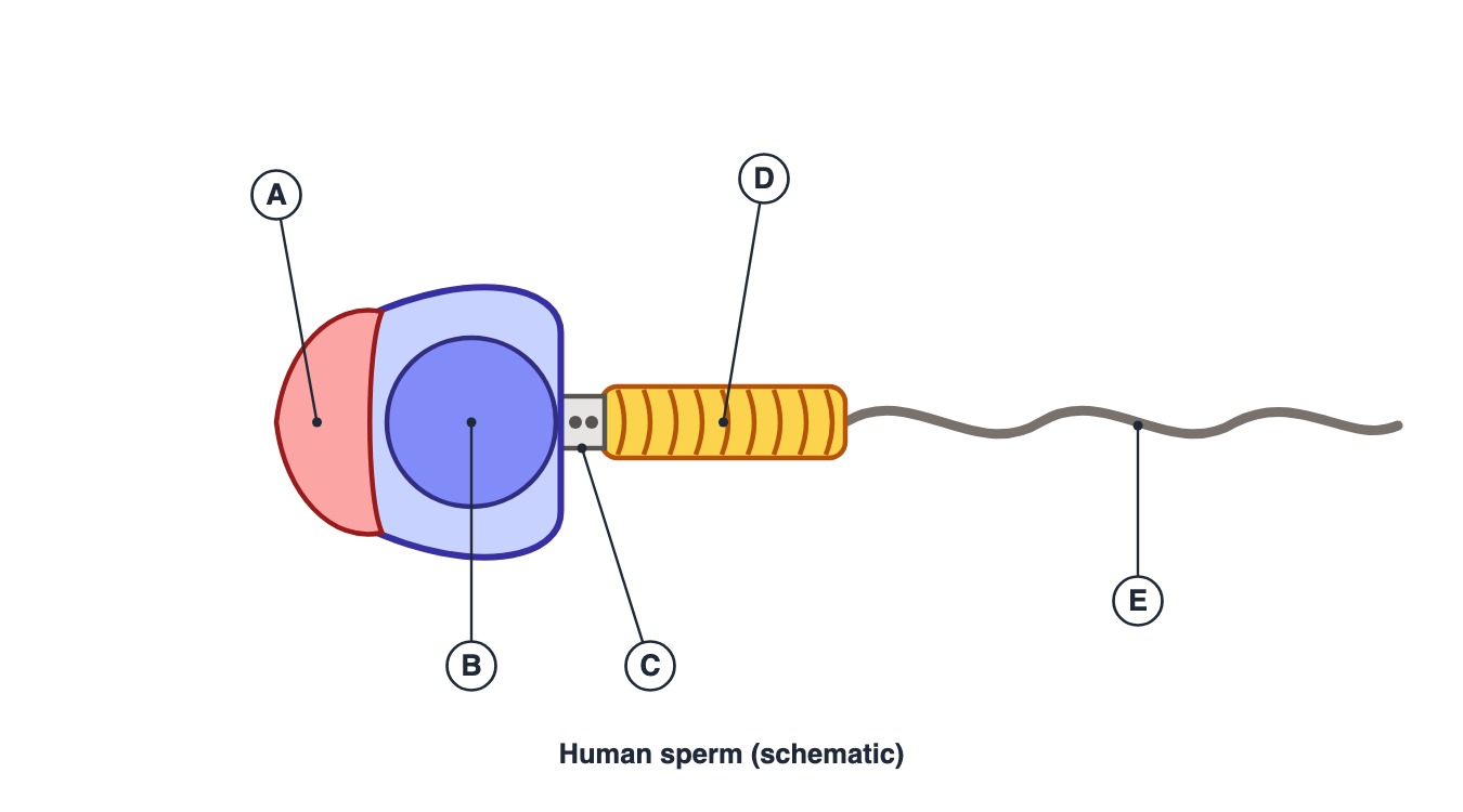
(a) middle piece
(b) centriole
(c) perforatorium of the tail
(d) acrosome
Show answer
Answer: (d) — The acrosome is a cap over the anterior part of the nucleus, formed from the Golgi complex, and contains enzymes such as hyaluronidase that dissolve the egg coverings during fertilisation.
Q14. Numerous mitochondria, which supply the energy for sperm motility, are present in the part marked
(a) E
(b) D
(c) C
(d) A
Show answer
Answer: (b) — D is the middle piece. It is packed with mitochondria that produce the ATP for the tail's movement.
Q15. The centrioles of the sperm are located in the part marked
(a) B
(b) E
(c) C
(d) D
Show answer
Answer: (c) — C is the neck, which contains the proximal and distal centrioles; the distal centriole gives rise to the axial filament of the tail.
NEET Previous Year Questions (PYQs)
Real NEET previous-year questions on this chapter, with explanations in our own words.
Q16. Which of the following statements is TRUE regarding Sertoli cells? (NEET PYQ)
(a) They provide nutrition to germ cells
(b) They are found outside the seminiferous tubule
(c) They synthesise androgens
(d) They form the acrosome
Show answer
Answer: (a) — Sertoli cells nourish the dividing germ cells inside the seminiferous tubule; Leydig cells (outside the tubule) make androgens.
Q17. The number of primary follicles present in each human ovary at puberty is approximately: (NEET PYQ)
(a) 6–10
(b) 60,000–80,000
(c) 2 million
(d) 400
Show answer
Answer: (b) — Most fetal follicles degenerate by puberty, leaving roughly 60,000–80,000 primary follicles per ovary.
Q18. FSH acts primarily on which cells to aid spermiogenesis? (NEET PYQ)
(a) Leydig cells
(b) Sertoli cells
(c) Spermatogonia directly
(d) Epididymal cells
Show answer
Answer: (b) — FSH acts on Sertoli cells, stimulating secretion of factors that help spermiogenesis.
Q19. The site of production of the male gamete is the: (NEET PYQ)
(a) Vas deferens
(b) Prostate
(c) Seminiferous tubule
(d) Epididymis
Show answer
Answer: (c) — Sperms are produced inside the seminiferous tubules of the testis.
Q20. A mature Graafian follicle releases the: (NEET PYQ)
(a) Ootid
(b) Secondary oocyte
(c) Zygote
(d) Primary oocyte
Show answer
Answer: (b) — Ovulation releases the secondary oocyte (commonly called the ovum), not the primary oocyte.
Q21. Which structure surrounds the secondary oocyte after it forms? (NEET PYQ)
(a) Chorionic villi
(b) Corpus luteum
(c) Zona pellucida
(d) Endometrium
Show answer
Answer: (c) — The zona pellucida is the new membrane that forms around the secondary oocyte.
Active Recall Prompt
Write everything you can recall about Human Reproduction (Part A), naming each part first: the male reproductive system (testis, scrotum, seminiferous tubule, Sertoli vs Leydig cells, sperm path, accessory glands); the female reproductive system (ovary, oviduct parts, uterine wall layers, mammary glands); spermatogenesis (spermatogonium → primary spermatocyte → secondary spermatocyte → spermatid → sperm, hormonal control, sperm structure); and oogenesis (oogonia, primary/secondary/tertiary/Graafian follicle, the unequal meiotic division, polar bodies). Begin each fact with its topic and end it with a full stop.