Kidney and Ureter for NEET PG Surgery: Renal and Ureteric Stones, Hydronephrosis, Haematuria, Renal Cell Carcinoma, Wilms Tumour and Renal Injury
Start Here
🎯 Exam priority: High. Expect: colic radiation by stone site, X-KUB + ultrasound and NCCT in acute colic (ICMR), 90% radio-opaque stones, stone composition, MET for stones under 10 mm, infected obstructed kidney needs drainage, PCNL for renal stones over 2 cm, haematuria work-up and cystoscopy age, RCC risks and nephrectomy, Wilms tumour and WAGR, renal trauma CT and nonoperative care, horseshoe kidney, PUJ obstruction, VUR and GU tuberculosis.
CBME mapping: SU29.1–29.6. Indian anchors: ICMR STWs on renal and ureteric stones (2025) and genitourinary TB (2022).
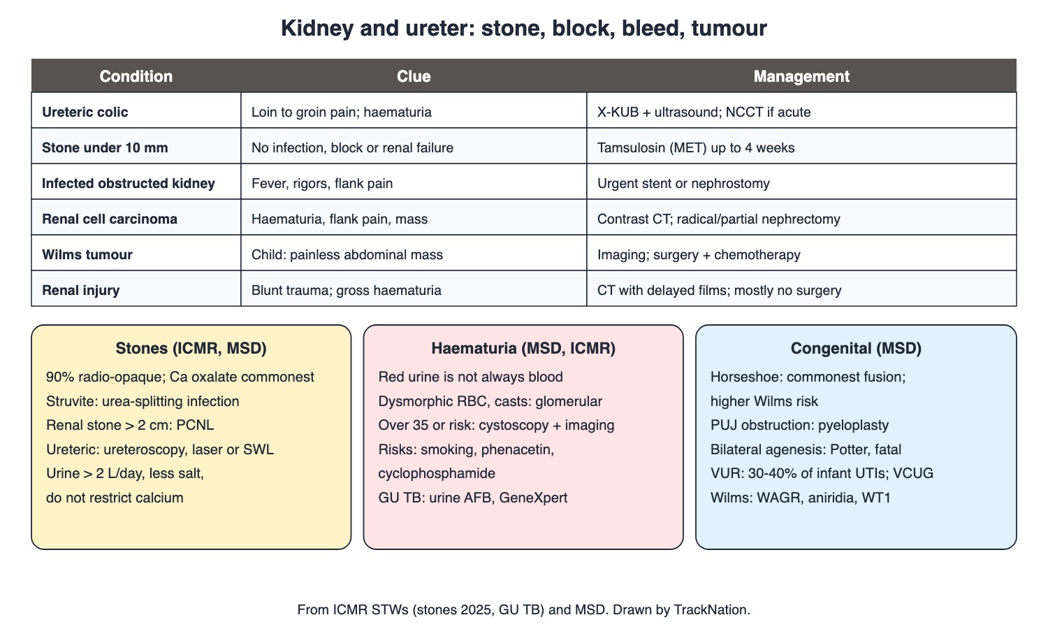
Kidney and ureter conditions at a glance. Drawn by TrackNation.
Renal and Ureteric Stones (ICMR, MSD)
Pain points to the site (ICMR): renal — flank; pelviureteric/upper ureter — back to front; mid ureter — back to groin; lower ureter — back to upper thigh and scrotum/vulva, sometimes voiding difficulty. Haematuria, nausea, vomiting; anuria with bilateral stones or a solitary kidney.
Imaging: X-KUB + ultrasound for all (90% radio-opaque; ultrasound safe in pregnancy, shows hydronephrosis); NCCT preferred in acute colic (MSD: preferred initial study); IVP if seen on X-ray; CT urography for radiolucent stones. Metabolic work-up for recurrent, bilateral, childhood, family-history or solitary-kidney cases.
Composition: calcium oxalate commonest; uric acid (urine pH <5.5, radiolucent); cystine (cystinuria); struvite from urea-splitting infection — can form staghorn calculi.
Treatment: analgesia (ketorolac, opioids); extra fluid does not speed passage. MET with an alpha-blocker (tamsulosin 0.4 mg) for ureteric stones <10 mm without infection, obstruction or renal failure, up to 4 weeks (MSD: unpassed by 6–8 weeks → removal). Warning signs needing referral: fever with rigors, anuria, renal failure, persistent haematuria.
Infection + obstruction → drain at once (ureteric stent or percutaneous nephrostomy). Removal: SWL; ureteroscopy with laser (mid/distal ureter); PCNL for renal stones >2 cm.
Prevention (ICMR): urine >2 L/day, less salt, more fibre and citrus; do not restrict calcium; cut oxalate (spinach, nuts) and purines. Recurrence is common.
Obstruction, Haematuria and GU TB (MSD, ICMR)
Obstructive uropathy: children — congenital (PUJ/VUJ narrowing, posterior urethral valves in boys); young adults — stones; older adults — BPH, cancers, strictures. Ultrasound first. Complete bladder/urethral block → anuria. Drain promptly if function falls, infection persists or pain is uncontrolled; immediately if infected.
Haematuria: red urine can be haemoglobin, myoglobin, porphyria, beets, drugs. Dysmorphic RBCs and RBC casts = glomerular. Genitourinary cancer mainly >50 or with risks (smoking, phenacetin, cyclophosphamide); cystoscopy and imaging for most patients >35 or with risk factors.
Genitourinary TB (ICMR): LUTS with dysuria/haematuria for ≥2 weeks not responding to antibiotics; early-morning urine AFB ×3–5 days, culture, GeneXpert; IVP/CT urography; 2HRZE/4HRE; watch for small contracted bladder and ureteric strictures.
Tumours, Trauma and Congenital (MSD)
Renal cell carcinoma (90–95% of renal cancers): risks smoking, obesity, hypertension, phenacetin, dialysis cysts, von Hippel-Lindau, tuberous sclerosis. Haematuria (commonest), flank pain, FUO, mass; often incidental; renal vein/IVC thrombus. Contrast CT/MRI; radical nephrectomy (with Gerota fascia) or partial nephrectomy.
Wilms tumour: embryonal kidney cancer of childhood; painless abdominal mass, sometimes haematuria or hypertension; WAGR (WT1 deletion, aniridia). Ultrasound/CT/MRI; surgery and chemotherapy, sometimes radiation.
Renal trauma: commonest GU injury; 85–90% blunt. Suspect high grade with gross haematuria or microscopic haematuria with hypotension → contrast CT with delayed images; stable microscopic haematuria alone needs no CT. Grades 1–5; most (all 1–2, most 3–4) managed non-operatively; intervene for persistent bleeding, expanding haematoma, pedicle avulsion.
Horseshoe kidney: commonest fusion anomaly; higher Wilms risk. PUJ obstruction → pyeloplasty. Bilateral agenesis (Potter) is fatal. VUR: 30–40% of infants with UTI; VCUG; mild grades resolve; scarring → hypertension.
Traps the Examiner Sets
Infected obstructed kidney: drain first, treat the stone later.
Don't restrict dietary calcium in stone formers.
Stable blunt trauma with microscopic haematuria alone needs no CT.
Uric acid stones are radiolucent.
Memory Hooks & Shortcuts
Shortcuts to lock in this article's numbers and lists. Each one only re-packages facts proven above.
🧠 Stone pain travels down with the stone: 'Back → front → groin → thigh' — colic
🧠 'Drain the pus before you chase the stone' — infected obstruction
🧠 'Uric acid hides on X-ray' — radiolucent
🧠 'Keep the milk, lose the salt' — prevention
🧠 Wilms: 'WAGR — Wilms, Aniridia, Genitourinary, intellectual disability' — syndrome
Rapid-Revision Cards
Lower ureteric stone
Pain to thigh, scrotum.
First tests
X-KUB + ultrasound.
Acute colic
NCCT.
Radio-opaque
90%.
Commonest stone
Calcium oxalate.
Staghorn
Struvite; urea-splitters.
Uric acid stone
pH <5.5; radiolucent.
MET
Tamsulosin; <10 mm; 4 weeks.
Infected + obstructed
Drain now.
Renal stone >2 cm
PCNL.
Mid-ureter
Ureteroscopy + laser.
Diet
>2 L urine; keep calcium.
Young adult obstruction
Stones.
PUV
Boys only.
Glomerular haematuria
Dysmorphic RBCs, casts.
Haematuria >35
Cystoscopy + imaging.
RCC
Haematuria; VHL; nephrectomy.
Wilms
Painless mass; WAGR.
Renal trauma
CT with delays; mostly no surgery.
Horseshoe
Commonest fusion.
GU TB
Urine AFB ×3–5; GeneXpert.
Sources
Facts in this article were checked against these documents.
Standard Treatment Workflows: Renal and Ureteric Stones (Nov 2025); Genitourinary Tuberculosis (2022) — ICMR / Department of Health Research, 2022–25
MSD Manual Professional: Urinary Calculi; Obstructive Uropathy; Hematuria; Renal Cell Carcinoma; Wilms Tumor; Renal Trauma; Kidney Anomalies; Vesicoureteral Reflux — Merck & Co. (MSD Manuals), 2025–26 (link)
Competency Based Undergraduate Curriculum, Vol III: General Surgery (SU29) — National Medical Commission, 2024
Test Yourself: Practice Questions
Thirty questions in NEET PG style, most of them clinical vignettes.
Practice Questions (NEET PG pattern)
Original questions written in the style of the exam: +4 for a correct answer, −1 for a wrong one. Every answer was checked against the sources listed above. NEET PG papers are not published by NBEMS, so these are not past-paper questions.
Q1. Colicky pain starting in the back and radiating to the upper thigh and scrotum suggests a stone in the
(a) renal pelvis
(b) lower ureter
(c) renal calyx
(d) upper ureter
Show answer
Answer: (b) — ICMR.
Q2. Per ICMR, every patient with suspected renal stones should first have
(a) retrograde pyelography
(b) renal biopsy
(c) X-ray KUB and ultrasound
(d) MRI
Show answer
Answer: (c) — NCCT preferred in acute colic.
Q3. The preferred imaging in acute renal colic, if available, is
(a) IVP
(b) MRI
(c) non-contrast CT
(d) plain X-ray only
Show answer
Answer: (c) — Detects radiolucent stones.
Q4. A pregnant woman with flank colic should be imaged first with
(a) CT
(b) IVP
(c) X-ray KUB with contrast
(d) ultrasound
Show answer
Answer: (d) — No radiation.
Q5. About what proportion of renal stones are radio-opaque?
(a) 50%
(b) 100%
(c) 90%
(d) 10%
Show answer
Answer: (c) — Uric acid stones are radiolucent.
Q6. The commonest type of urinary stone is
(a) uric acid
(b) struvite
(c) calcium oxalate
(d) cystine
Show answer
Answer: (c) — 40–80%.
Q7. A staghorn calculus in a woman with recurrent UTIs from urea-splitting bacteria is most likely
(a) cystine
(b) calcium oxalate monohydrate
(c) struvite (magnesium ammonium phosphate)
(d) uric acid
Show answer
Answer: (c) — Urea-splitting bacteria.
Q8. Uric acid stones form in urine with
(a) pH below 5.5
(b) urea-splitting bacteria
(c) high citrate
(d) pH above 8
Show answer
Answer: (a) — Radiolucent.
Q9. A patient has a 6 mm lower ureteric stone, controlled pain, no fever and normal renal function. Per ICMR, offer
(a) immediate PCNL
(b) IV fluids to flush it
(c) medical expulsive therapy with tamsulosin for up to 4 weeks
(d) open ureterolithotomy
Show answer
Answer: (c) — Alpha-blocker.
Q10. Forcing extra IV fluids in renal colic
(a) dissolves calcium stones
(b) doubles passage rate
(c) is mandatory
(d) does not speed stone passage
Show answer
Answer: (d) — MSD.
Q11. A patient with a ureteric stone has fever with rigors and a hydronephrotic kidney. Immediate priority:
(a) drain the kidney (stent or nephrostomy) with antibiotics
(b) observe 4 weeks
(c) SWL today
(d) medical expulsive therapy
Show answer
Answer: (a) — Infected obstruction.
Q12. The treatment of choice for a 3 cm renal pelvic stone is
(a) SWL alone always
(b) medical expulsive therapy
(c) observation
(d) percutaneous nephrolithotomy
Show answer
Answer: (d) — Renal stones >2 cm.
Q13. A mid-ureteric stone that fails to pass is usually treated by
(a) PCNL
(b) ureteroscopy with laser lithotripsy
(c) open pyelolithotomy
(d) nephrectomy
Show answer
Answer: (b) — SWL alternative.
Q14. Dietary advice to a calcium-stone former per ICMR includes
(a) high salt
(b) strict calcium restriction
(c) more spinach
(d) urine over 2 L/day and do not restrict calcium
Show answer
Answer: (d) — Reduce oxalate, salt.
Q15. The commonest cause of urinary obstruction in young adults is
(a) posterior urethral valves
(b) BPH
(c) prostate cancer
(d) calculi
Show answer
Answer: (d) — Older adults: BPH.
Q16. Posterior urethral valves occur in
(a) adults only
(b) girls only
(c) boys only
(d) both sexes equally
Show answer
Answer: (c) — Congenital obstruction.
Q17. Red urine with a negative microscopy for RBCs may be due to
(a) stones
(b) renal cell carcinoma
(c) myoglobin, haemoglobin, beets or drugs
(d) bladder cancer
Show answer
Answer: (c) — Not haematuria.
Q18. Haematuria with dysmorphic RBCs and RBC casts points to a
(a) ureteric stone
(b) urethral injury
(c) bladder tumour
(d) glomerular cause
Show answer
Answer: (d) — Nephrology work-up.
Q19. A 60-year-old smoker has painless visible haematuria. Besides imaging, he needs
(a) reassurance
(b) antibiotics only
(c) renal biopsy first
(d) cystoscopy
Show answer
Answer: (d) — Over 35 or risk factors.
Q20. The commonest presenting feature of renal cell carcinoma is
(a) bone pain
(b) polycythaemia
(c) varicocele
(d) haematuria
Show answer
Answer: (d) — Often found incidentally.
Q21. A genetic syndrome strongly linked to renal cell carcinoma is
(a) Down syndrome
(b) Marfan syndrome
(c) von Hippel-Lindau disease
(d) Turner syndrome
Show answer
Answer: (c) — Also tuberous sclerosis.
Q22. Standard treatment for localised renal cell carcinoma is
(a) chemotherapy alone
(b) radiotherapy alone
(c) observation
(d) radical or partial nephrectomy
Show answer
Answer: (d) — Gerota fascia removed.
Q23. A 3-year-old has a painless abdominal mass. Most likely
(a) renal cell carcinoma
(b) horseshoe kidney
(c) hydatid cyst
(d) Wilms tumour
Show answer
Answer: (d) — Embryonal.
Q24. WAGR syndrome combines Wilms tumour with
(a) aniridia, genitourinary anomalies and intellectual disability
(b) hemihypertrophy only
(c) deafness and cataract
(d) polydactyly
Show answer
Answer: (a) — WT1 deletion.
Q25. After a fall, a stable patient has gross haematuria. The investigation is
(a) no imaging
(b) contrast CT with delayed images
(c) cystoscopy
(d) IVP only
Show answer
Answer: (b) — High-grade suspicion.
Q26. A haemodynamically stable blunt-trauma patient with only microscopic haematuria
(a) needs angiography
(b) needs urgent nephrectomy
(c) usually has a minor injury and needs no CT
(d) needs cystoscopy
Show answer
Answer: (c) — MSD.
Q27. Most renal injuries, including most grade 3 and 4 injuries, are managed
(a) by stent in all
(b) by open repair always
(c) by nephrectomy
(d) non-operatively
Show answer
Answer: (d) — Intervene for persistent bleeding.
Q28. The commonest renal fusion anomaly, which also carries a higher Wilms risk, is
(a) duplex kidney
(b) crossed fused ectopia
(c) pancake kidney
(d) horseshoe kidney
Show answer
Answer: (d) — Lower poles join.
Q29. An infant with a febrile UTI is found to have VUR. The key diagnostic study is
(a) IVP
(b) DMSA alone
(c) voiding cystourethrogram
(d) cystoscopy
Show answer
Answer: (c) — 30–40% of infant UTIs.
Q30. A man has dysuria and haematuria for over 2 weeks despite antibiotics. Per ICMR, send
(a) urine cytology only
(b) PSA
(c) early-morning urine for AFB and GeneXpert
(d) serum calcium
Show answer
Answer: (c) — Genitourinary TB.
Active Recall
From memory: describe the presentation, imaging and management of renal and ureteric stones including MET and infected obstruction, outline the haematuria work-up, describe renal cell carcinoma, Wilms tumour and renal trauma, and summarise congenital anomalies, VUR and genitourinary TB.