Bacterial Cell, Growth and Classification for NEET PG Microbiology: Gram-positive vs Gram-negative Walls, Spores, Flagella, Oxygen Needs and History
Start Here
🎯 Exam priority: Moderate–high. Expect: who did what in early microbiology, differences between gram-positive and gram-negative walls, endotoxin vs exotoxin, spore-formers, flagella arrangement examples, oxygen classes with examples, growth-curve phases, generation times, and virulence factors such as capsules, IgA protease, biofilm and superantigens.
CBME mapping: MI1.1–1.2. Fact-checked against OpenStax Microbiology and the MSD Manual.
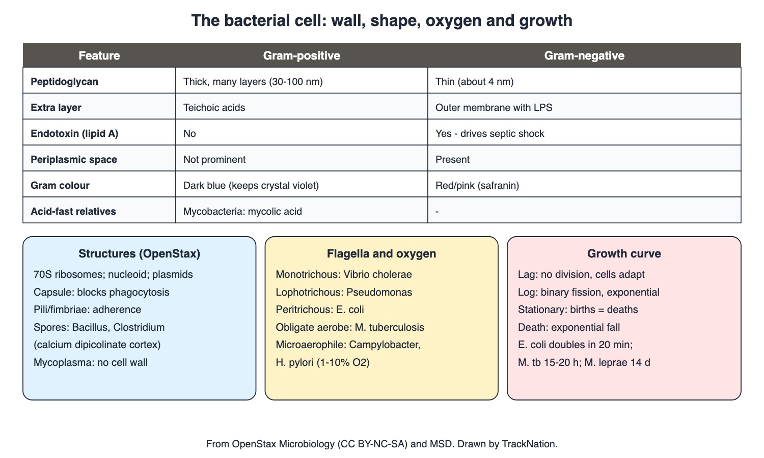
Cell wall types, structures, flagella, oxygen classes and the growth curve. Drawn by TrackNation.
Pioneers (OpenStax)
Leeuwenhoek (1675): first saw single-celled "animalcules" with his simple microscope.
Pasteur: fermentation by microbes (1856); swan-neck flasks disproved spontaneous generation — foundation of the germ theory.
Semmelweis: handwashing with chlorinated lime water cut puerperal fever deaths. John Snow (1848): traced cholera to sewage-contaminated drinking water — the first known epidemiological study.
Lister (1867): carbolic acid (phenol) spray in surgery — antiseptic surgery. Koch: postulates linking one microbe to one disease; identified the causes of anthrax, tuberculosis and cholera.
The Bacterial Cell (OpenStax, MSD)
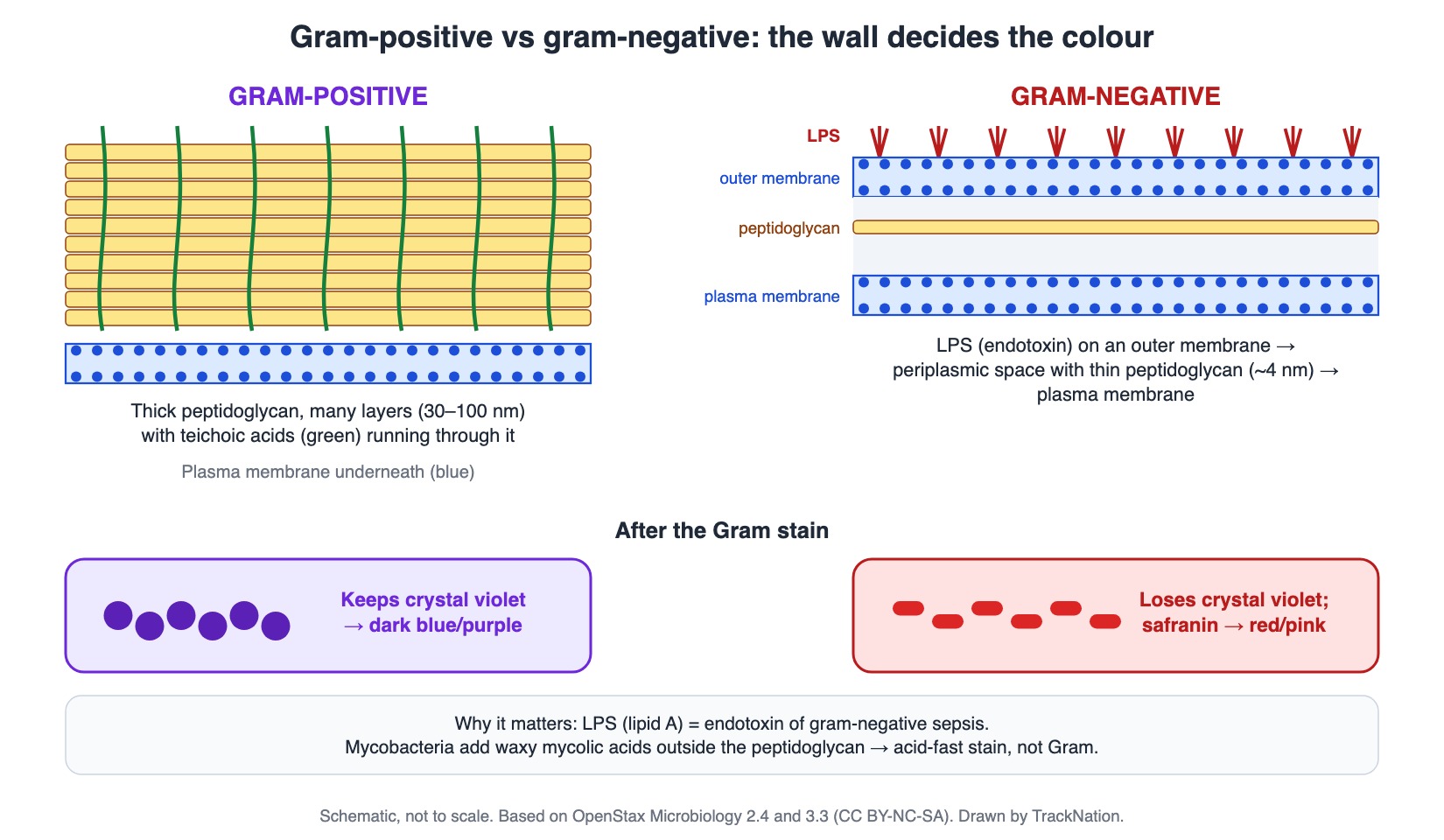
Gram-positive vs gram-negative walls and why they stain differently. Drawn by TrackNation.
Bacteria have circular double-stranded DNA (nucleoid), 70S ribosomes (eukaryotes 80S), often plasmids carrying useful genes such as resistance, and a cell wall — except Mycoplasma.
Peptidoglycan (murein) — alternating NAG and NAM chains — exists only in bacteria. Gram-positive: thick (30–100 nm) layers with teichoic acids; gram-negative: thin (~4 nm) layer, periplasmic space and an outer membrane with lipopolysaccharide (endotoxin).
Gram stain: crystal violet → iodine → alcohol → safranin; gram-positive stays dark blue, gram-negative turns red. Mycobacteria have waxy mycolic acids — acid-fast (Ziehl–Neelsen, Kinyoun); acid-fast staining also picks up Nocardia and Cryptosporidium, Cyclospora, Cystoisospora.
Endospores (calcium dipicolinate cortex) — Bacillus and Clostridium (anthrax, tetanus, gas gangrene, botulism).
Flagella: monotrichous — Vibrio cholerae; lophotrichous (tuft at one end) — Pseudomonas aeruginosa; amphitrichous — both ends; peritrichous — E. coli. Pili/fimbriae mediate adherence.
Oxygen, Temperature and Growth (OpenStax, MSD)
Obligate aerobes: M. tuberculosis. Obligate anaerobes: killed by oxygen (Bacteroides, Clostridium). Facultative: grow with or without O2 (E. coli, S. aureus). Microaerophiles: need low oxygen (~1–10%) — Campylobacter, H. pylori. Obligate intracellular: chlamydiae, rickettsiae.
Oxygen-tolerant organisms detoxify ROS with superoxide dismutase, peroxidase and catalase.
Human pathogens are mesophiles (optimum ~20–45 °C); psychrophiles grow at 0 °C or below; thermophiles 50–80 °C.
Growth curve: lag (no division, cells adapt) → log (binary fission, exponential) → stationary (new = dying) → death. Generation time: as little as 20 minutes (E. coli), 15–20 hours (M. tuberculosis), 14 days (M. leprae). Slow-metabolising persisters survive antibiotics.
Virulence Factors (MSD)
Capsule (pneumococcus, meningococcus, Hib) blocks phagocytosis; capsule-specific antibodies opsonise.
Enzymes (hyaluronidase, collagenase, elastase) spread infection; IgA protease (gonococcus, meningococcus, H. influenzae, pneumococcus) destroys mucosal secretory IgA.
Exotoxins are proteins (diphtheria, cholera, tetanus, botulism); some are preformed in food (botulism, staphylococcal and Bacillus cereus food poisoning). Endotoxin = LPS of gram-negative outer membrane — activates complement, clotting and kinins; drives gram-negative septic shock.
Superantigens activate huge numbers of T cells — toxic shock syndrome (staphylococcal, streptococcal). Biofilm (Pseudomonas in cystic fibrosis; staphylococci on catheters and prostheses) resists phagocytes and antibiotics. Legionella and Listeria evade the oxidative burst.
Traps the Examiner Sets
Mycoplasma has no cell wall, so it cannot be seen on a Gram stain.
Endotoxin is lipid A of gram-negative LPS; exotoxins are proteins from either group.
Campylobacter and H. pylori are microaerophiles; M. tuberculosis is an obligate aerobe.
Pseudomonas is lophotrichous; Vibrio monotrichous; E. coli peritrichous.
Memory Hooks & Shortcuts
Shortcuts to lock in this article's numbers and lists. Each one only re-packages facts proven above.
🧠 History chain: 'Leeuwenhoek Looked, Pasteur Proved, Semmelweis Scrubbed, Snow Mapped, Lister Sprayed, Koch Confirmed' — history
🧠 'Gram-Positive keeps the violet (dark blue) in its Plenty of Peptidoglycan; gram-Negative loses it and turns red' — Gram stain
🧠 Flagella: 'Vibrio one, Pseudomonas tuft, E. coli all over' — flagella
🧠 'Cam and Helico like it Low' — microaerophiles — oxygen
🧠 Doubling times '20 minutes – 20 hours – 2 weeks': E. coli, M. tb, M. leprae — growth
Rapid-Revision Cards
Leeuwenhoek
Animalcules, 1675.
Pasteur
Swan-neck flasks; fermentation.
Semmelweis
Handwashing (chlorinated lime).
Snow
Cholera via water; 1st epidemiology.
Lister
Phenol, 1867.
Koch
Postulates; anthrax, TB, cholera.
Peptidoglycan
NAG + NAM; bacteria only.
Gram +
Thick wall, teichoic acid.
Gram −
Thin wall, outer membrane, LPS.
Gram colours
+ dark blue; − red.
Acid-fast
Mycolic acid; Nocardia, Cryptosporidium.
No cell wall
Mycoplasma.
Ribosomes
70S (eukaryote 80S).
Spore-formers
Bacillus, Clostridium.
Flagella
Vibrio mono; Pseudomonas lopho; E. coli peri.
Microaerophiles
Campylobacter, H. pylori.
Obligate intracellular
Chlamydia, Rickettsia.
Growth curve
Lag → log → stationary → death.
Doubling
E. coli 20 min; M. tb 15–20 h; M. leprae 14 d.
Endotoxin
LPS; gram-negative sepsis.
Superantigen
Toxic shock syndrome.
Sources
Facts in this article were checked against these documents.
Microbiology (sections 1.1, 3.1, 3.2, 3.3, 9.1, 9.2, 9.4) — Parker N, Schneegurt M, Thi Tu A-H, Lister P, Forster BM (OpenStax), 2016 (link)
MSD Manual Professional: Overview of Bacteria; Factors Facilitating Microbial Invasion — Merck & Co. (MSD Manuals), 2024–25 (link)
Competency Based Undergraduate Curriculum, Vol I: Microbiology (MI1.1–1.2) — National Medical Commission, 2024
Test Yourself: Practice Questions
Thirty questions in NEET PG style, most of them clinical vignettes.
Practice Questions (NEET PG pattern)
Original questions written in the style of the exam: +4 for a correct answer, −1 for a wrong one. Every answer was checked against the sources listed above. NEET PG papers are not published by NBEMS, so these are not past-paper questions.
Q1. The first person to see single-celled 'animalcules' with a microscope (1675) was
(a) Joseph Lister
(b) Robert Koch
(c) Louis Pasteur
(d) Antonie van Leeuwenhoek
Show answer
Answer: (d) — OpenStax.
Q2. Spontaneous generation was disproved by experiments with
(a) Koch's postulates
(b) Pasteur's swan-neck flasks
(c) Gram's stain
(d) Lister's phenol spray
Show answer
Answer: (b) — Airborne microbes trapped.
Q3. Semmelweis reduced puerperal fever deaths by
(a) vaccination
(b) phenol spray
(c) boiling instruments
(d) handwashing with chlorinated lime water
Show answer
Answer: (d) — OpenStax.
Q4. John Snow's 1848 London work showed that cholera spreads through
(a) air (miasma)
(b) rats
(c) mosquitoes
(d) contaminated drinking water
Show answer
Answer: (d) — First epidemiological study.
Q5. Lister introduced antiseptic surgery in 1867 using
(a) chlorine gas
(b) carbolic acid (phenol)
(c) iodine
(d) alcohol
Show answer
Answer: (b) — OpenStax.
Q6. Koch's postulates link
(a) a specific microbe to a specific disease
(b) genes to traits
(c) fermentation to spoilage
(d) vaccines to immunity
Show answer
Answer: (a) — Anthrax, TB, cholera.
Q7. Peptidoglycan is made of alternating
(a) glucose and galactose
(b) teichoic and lipoteichoic acids
(c) lipid A and O-antigen
(d) N-acetylglucosamine and N-acetylmuramic acid
Show answer
Answer: (d) — Only in bacteria.
Q8. A feature of the gram-positive wall is
(a) endotoxin
(b) an outer membrane with LPS
(c) a prominent periplasmic space
(d) thick peptidoglycan with teichoic acids
Show answer
Answer: (d) — 30–100 nm.
Q9. The gram-negative outer membrane contains
(a) chitin
(b) teichoic acid
(c) mycolic acid
(d) lipopolysaccharide (endotoxin)
Show answer
Answer: (d) — Lipid A.
Q10. In the Gram stain, gram-negative bacteria appear red because they
(a) retain crystal violet
(b) lose crystal violet on alcohol decolourisation and take up safranin
(c) are acid-fast
(d) lack DNA
Show answer
Answer: (b) — MSD.
Q11. Mycobacteria resist Gram staining because their wall contains
(a) waxy mycolic acids
(b) teichoic acids
(c) LPS
(d) no peptidoglycan
Show answer
Answer: (a) — Acid-fast stains.
Q12. Besides mycobacteria, acid-fast staining identifies
(a) Nocardia and Cryptosporidium
(b) Candida
(c) E. coli
(d) Staphylococcus
Show answer
Answer: (a) — Also Cyclospora, Cystoisospora.
Q13. A bacterium with no cell wall, hence not seen on Gram stain, is
(a) Streptomyces
(b) Mycoplasma
(c) Clostridium
(d) Vibrio
Show answer
Answer: (b) — MSD.
Q14. Bacterial ribosomes are
(a) 90S
(b) 70S
(c) 60S
(d) 80S
Show answer
Answer: (b) — Eukaryotic 80S.
Q15. Clinically important endospore-forming genera are
(a) Bacillus and Clostridium
(b) Neisseria and Moraxella
(c) Vibrio and Campylobacter
(d) Staphylococcus and Streptococcus
Show answer
Answer: (a) — Gram-positive.
Q16. The endospore cortex contains
(a) LPS
(b) hyaluronic acid
(c) teichoic acid
(d) calcium dipicolinate
Show answer
Answer: (d) — OpenStax.
Q17. A single polar flagellum (monotrichous) is characteristic of
(a) Salmonella
(b) E. coli
(c) Vibrio cholerae
(d) Proteus
Show answer
Answer: (c) — OpenStax.
Q18. Pseudomonas aeruginosa has flagella arranged as
(a) lophotrichous (tuft at one end)
(b) amphitrichous
(c) none
(d) peritrichous
Show answer
Answer: (a) — OpenStax.
Q19. E. coli flagella are
(a) absent
(b) lophotrichous
(c) peritrichous
(d) monotrichous
Show answer
Answer: (c) — OpenStax.
Q20. Campylobacter and H. pylori are
(a) obligate anaerobes
(b) aerotolerant anaerobes
(c) microaerophiles
(d) obligate aerobes
Show answer
Answer: (c) — Low O2.
Q21. An obligate aerobe among human pathogens is
(a) Mycobacterium tuberculosis
(b) Clostridium tetani
(c) Bacteroides fragilis
(d) Campylobacter jejuni
Show answer
Answer: (a) — OpenStax.
Q22. Obligate intracellular bacteria include
(a) S. aureus and S. pyogenes
(b) Bacillus and Clostridium
(c) chlamydiae and rickettsiae
(d) E. coli and Klebsiella
Show answer
Answer: (c) — MSD.
Q23. Enzymes that protect bacteria from reactive oxygen species include
(a) beta-lactamase
(b) coagulase and DNase
(c) urease and oxidase
(d) superoxide dismutase, peroxidase and catalase
Show answer
Answer: (d) — OpenStax.
Q24. In the bacterial growth curve, cell numbers stay constant while cells adapt and enlarge in the
(a) lag phase
(b) stationary phase
(c) death phase
(d) log phase
Show answer
Answer: (a) — OpenStax.
Q25. In the stationary phase
(a) all cells die
(b) cells divide fastest
(c) new cells equal dying cells
(d) no metabolism occurs
Show answer
Answer: (c) — Plateau.
Q26. The generation time of M. tuberculosis is about
(a) 1 hour
(b) 14 days
(c) 20 minutes
(d) 15–20 hours
Show answer
Answer: (d) — M. leprae 14 days.
Q27. Encapsulated bacteria such as pneumococcus resist host defence mainly by
(a) producing catalase
(b) forming spores
(c) producing endotoxin
(d) blocking phagocytosis
Show answer
Answer: (d) — MSD.
Q28. Gonococci and meningococci evade mucosal immunity by producing
(a) coagulase
(b) urease
(c) streptokinase
(d) IgA protease
Show answer
Answer: (d) — MSD.
Q29. Toxic shock syndrome results from
(a) capsule
(b) superantigens activating large numbers of T cells
(c) endotoxin from gram-positive cocci
(d) IgE-mediated reaction
Show answer
Answer: (b) — Staph/strep.
Q30. Infection of an IV catheter by staphylococci is hard to clear because of
(a) biofilm
(b) mycolic acid
(c) spores
(d) flagella
Show answer
Answer: (a) — Resists phagocytes and antibiotics.
Active Recall
From memory: name the key pioneers and their contributions, compare gram-positive and gram-negative walls, list bacterial structures with examples (spores, flagella, capsule), classify bacteria by oxygen need, describe the growth curve and generation times, and explain the main virulence factors.