Specimens, Stains, Culture Media and Susceptibility Testing for NEET PG Microbiology: Gram, ZN, India Ink, Selective Media, MIC and Kirby-Bauer
Start Here
🎯 Exam priority: High. Expect: "which medium for which organism" (TCBS, Thayer-Martin, BCYE, Lowenstein-Jensen, Tinsdale, Bordet-Gengou), the order of reagents in Gram and Ziehl–Neelsen staining, which stain for which organism (India ink, KOH, Giemsa, silver, auramine), blood-culture technique, and MIC vs disk diffusion.
CBME mapping: MI1.4–1.6, MI1.10, MI11.1. Fact-checked against OpenStax Microbiology and the MSD Manual.
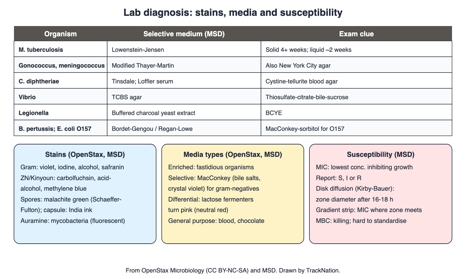
Selective media by organism, stains, media types and susceptibility tests. Drawn by TrackNation.
Getting the Specimen Right (MSD)
Culture is the gold standard for identification but may take days or weeks; transport specimens quickly, in the correct medium, and refrigerate if delayed (in most cases) so contaminating flora do not overgrow.
Swabs matter: cotton-tipped swabs are toxic to some bacteria, including chlamydiae; wooden-shafted swabs are toxic to some viruses.
Blood cultures: scrub skin with 70% isopropyl alcohol plus chlorhexidine and/or iodine; take multiple samples from different sites, ideally near fever spikes. Skin flora growing in only one bottle is usually contamination. Moulds such as Aspergillus usually do not grow from blood.
Anaerobes: never culture from sites where they are normal flora; shield the specimen from air (anaerobic transport media).
Mycobacteria: sputum is decontaminated (NaOH) and concentrated; liquid automated systems grow M. tuberculosis in about 2 weeks vs ≥4 weeks on Lowenstein–Jensen; keep cultures 8 weeks before calling them negative (M. ulcerans: up to 12 weeks at 32 °C).
Viral culture has largely been replaced by nucleic acid tests for common viruses.
Stains (OpenStax 2.4, MSD)
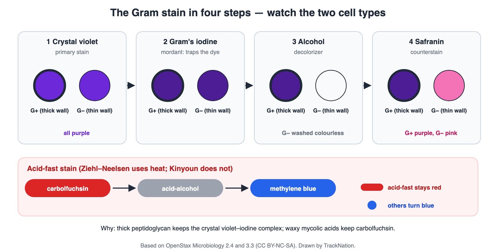
The Gram stain step by step, and the acid-fast stain. Drawn by TrackNation.
Fixation (heat or chemical) attaches and kills cells. Basic (positive) dyes: crystal violet, methylene blue, safranin, malachite green, basic fuchsin. Acidic dyes (eosin, acid fuchsin, rose bengal) are repelled by the negatively charged cell wall — the basis of negative staining.
Gram: crystal violet → iodine → decolouriser (ethanol or acetone–ethanol) → safranin; gram-positive purple/blue, gram-negative red. It also shows shape, arrangement and pus cells (bacterial infection). Microscopy needs about 104–105 organisms/mL.
Acid-fast (Ziehl–Neelsen uses heat, Kinyoun does not): carbolfuchsin → acid-alcohol → methylene blue; acid-fast organisms stay red. Sputum smear needs ≥10,000 bacilli/mL. Modified acid-fast stains pick up Nocardia and oocysts of Cryptosporidium, Cystoisospora, Cyclospora; Wade–Fite (modified ZN) for M. leprae. Auramine–rhodamine (fluorescent) for mycobacteria; calcofluor white for fungi.
Spores: Schaeffer–Fulton — malachite green driven in by heat, safranin counterstain. Capsule: negative staining with India ink or nigrosin — Cryptococcus neoformans in CSF shows a halo (less sensitive than cryptococcal antigen).
Wet mount / dark-field: fungi, parasites, clue cells (bacterial vaginosis), motile Trichomonas, Treponema. 10% KOH dissolves tissue to show fungi.
Wright–Giemsa: blood parasites, Histoplasma in phagocytes, viral and chlamydial inclusions, Pneumocystis trophozoites. Silver stains (Warthin–Starry, Dieterle): spirochaetes, H. pylori, Bartonella henselae. Trichrome: intestinal protozoa. Direct fluorescent antibody tests: Pneumocystis, Legionella.
Culture Media (OpenStax 9.6, MSD)
Chemically defined (exact composition known) vs complex (extracts and digests). Enriched media add growth factors for fastidious organisms. General-purpose plates: blood and chocolate agar.
Selective: MacConkey — bile salts and crystal violet suppress gram-positives. Differential: on MacConkey, lactose fermenters turn pink (neutral red indicator); mannitol salt agar turns yellow with S. aureus.
Selective media by organism (MSD): M. tuberculosis — Lowenstein–Jensen; N. gonorrhoeae / N. meningitidis — modified Thayer–Martin (or New York City); C. diphtheriae — Tinsdale, cystine-tellurite blood agar, Löffler serum; B. pertussis — Bordet–Gengou or Regan–Lowe; Legionella — buffered charcoal yeast extract (BCYE); Vibrio — TCBS; Campylobacter — cefoperazone-vancomycin selective agars; Yersinia — CIN; E. coli O157 — MacConkey-sorbitol; Salmonella/Shigella — MacConkey or EMB, alternatively XLD, Hektoen, SS agar or selenite enrichment broth.
Antimicrobial Susceptibility Testing (MSD)
MIC = the lowest concentration that inhibits growth in vitro, translated to S, I or R (sometimes "non-susceptible"). Used for bacteria including mycobacteria and anaerobes, and some fungi (Candida).
MBC (minimal bactericidal concentration) shows whether a drug is bacteriostatic or bactericidal, but is technically difficult and lacks agreed standards.
Disk diffusion (Kirby–Bauer) for rapidly growing organisms: antibiotic disks on an inoculated plate; after 16–18 hours the zone of inhibition diameter is measured (qualitative S/I/R).
Gradient strip: a film carrying an antibiotic gradient — the MIC is read where inhibition begins on the strip. Synergy testing checks drug combinations. Nucleic acid tests detect resistance genes such as mecA (oxacillin/methicillin resistance in S. aureus).
Traps the Examiner Sets
Chlamydia swab: avoid cotton-tipped swabs (toxic).
One bottle of coagulase-negative skin flora = usually contamination; multiple bottles from different sites are needed.
Ziehl–Neelsen decolouriser is acid-alcohol; Gram decolouriser is alcohol/acetone.
Kirby–Bauer gives a zone diameter (category), not the MIC; the gradient strip gives an MIC.
India ink is less sensitive than cryptococcal antigen for cryptococcal meningitis.
Memory Hooks & Shortcuts
Shortcuts to lock in this article's numbers and lists. Each one only re-packages facts proven above.
🧠 'Very Cold Lemon Juice': TCBS for Vibrio, Charcoal (BCYE) for Legionella, LJ for M. tuberculosis — and 'Thayer for the Two Neisseria' — media
🧠 Gram order 'Come In And Stain': Crystal violet, Iodine, Alcohol, Safranin — Gram stain
🧠 ZN 'Carbolfuchsin – Acid – Methylene blue': 'Can AFB Mask?' — acid-fast
🧠 'India ink shows the Invisible capsule' — negative stain, Cryptococcus halo — capsule
🧠 'MIC is a number, Kirby is a circle' — strip gives MIC, disk gives zone — AST
Rapid-Revision Cards
Gold standard
Culture (slow).
Chlamydia swab
No cotton tip (toxic).
Blood culture prep
70% isopropyl + chlorhexidine/iodine.
One bottle skin flora
Contamination.
Mycobacterial culture
NaOH decontaminate; hold 8 weeks.
Gram decolouriser
Ethanol or acetone–ethanol.
ZN
Carbolfuchsin, acid-alcohol, methylene blue.
Smear threshold
≥10,000 AFB/mL.
Modified AFB
Nocardia; Cryptosporidium, Cystoisospora, Cyclospora.
Auramine
Fluorescent; mycobacteria.
Spore stain
Malachite green (Schaeffer–Fulton).
India ink
Cryptococcus halo; antigen better.
KOH 10%
Fungi in tissue.
Dark-field
Treponema; clue cells; Trichomonas.
Silver stain
H. pylori, Bartonella, spirochaetes.
MacConkey
Bile salts + crystal violet; lactose = pink.
TCBS / BCYE
Vibrio / Legionella.
Thayer–Martin
Gonococcus, meningococcus.
Tinsdale, Löffler
C. diphtheriae.
MIC
Lowest inhibitory conc.; gradient strip.
Kirby–Bauer
Zone diameter, 16–18 h.
Sources
Facts in this article were checked against these documents.
Microbiology (sections 2.4, 9.6) — Parker N, Schneegurt M, Thi Tu A-H, Lister P, Forster BM (OpenStax), 2016 (link)
MSD Manual Professional: Culture; Microscopy; Susceptibility Testing; Introduction to Laboratory Diagnosis of Infectious Disease — Merck & Co. (MSD Manuals), 2024–25 (link)
Competency Based Undergraduate Curriculum, Vol I: Microbiology (MI1.4–1.6, MI1.10, MI11.1) — National Medical Commission, 2024
Test Yourself: Practice Questions
Thirty questions in NEET PG style, many of them bedside or bench scenarios.
Practice Questions (NEET PG pattern)
Original questions written in the style of the exam: +4 for a correct answer, −1 for a wrong one. Every answer was checked against the sources listed above. NEET PG papers are not published by NBEMS, so these are not past-paper questions.
Q1. The gold standard for identifying most bacterial pathogens, despite taking days, is
(a) serology
(b) MALDI-TOF on the direct specimen
(c) culture
(d) Gram stain
Show answer
Answer: (c) — MSD.
Q2. A urethral swab for Chlamydia trachomatis should not be collected with a cotton-tipped swab because cotton
(a) contains DNA
(b) is toxic to chlamydiae
(c) absorbs too much fluid
(d) causes false positives
Show answer
Answer: (b) — MSD.
Q3. Coagulase-negative staphylococci grow in only one of three blood-culture sets from a febrile patient without a device. This most likely represents
(a) laboratory error in identification
(b) endocarditis
(c) skin contamination
(d) true bacteraemia
Show answer
Answer: (c) — Single bottle of skin flora.
Q4. Skin preparation recommended before drawing blood cultures is
(a) no preparation
(b) 70% isopropyl alcohol with chlorhexidine and/or tincture of iodine
(c) povidone-iodine after the draw
(d) soap and water only
Show answer
Answer: (b) — MSD.
Q5. A fungus that usually cannot be grown from blood cultures is
(a) Candida albicans
(b) Cryptococcus
(c) Aspergillus
(d) Histoplasma
Show answer
Answer: (c) — Moulds.
Q6. Sputum for mycobacterial culture is first
(a) heat-fixed
(b) stored at room temperature for 48 h
(c) decontaminated with NaOH and concentrated
(d) diluted in saline
Show answer
Answer: (c) — Kills normal flora.
Q7. Before a mycobacterial culture is reported negative, it should be incubated for
(a) 4 days
(b) 2 weeks
(c) 8 weeks
(d) 6 months
Show answer
Answer: (c) — MSD.
Q8. In the Gram stain, the decolourising step uses
(a) acid-alcohol
(b) ethanol or acetone–ethanol
(c) water
(d) safranin
Show answer
Answer: (b) — ZN uses acid-alcohol.
Q9. The primary stain in both Ziehl–Neelsen and Kinyoun methods is
(a) carbolfuchsin
(b) malachite green
(c) methylene blue
(d) crystal violet
Show answer
Answer: (a) — Counterstain methylene blue.
Q10. Detection of M. tuberculosis on a sputum smear requires at least about
(a) 100 organisms/mL
(b) 10,000 organisms/mL
(c) 10 organisms/mL
(d) 1 million organisms/mL
Show answer
Answer: (b) — Limits sensitivity.
Q11. A modified acid-fast stain of stool from an HIV patient with chronic diarrhoea is used to detect
(a) Cryptosporidium oocysts
(b) Entamoeba cysts
(c) Strongyloides larvae
(d) Giardia trophozoites
Show answer
Answer: (a) — Also Cystoisospora, Cyclospora.
Q12. A fluorescent stain used to screen for mycobacteria is
(a) auramine–rhodamine
(b) calcofluor white
(c) acridine orange only
(d) Giemsa
Show answer
Answer: (a) — Calcofluor for fungi.
Q13. In the Schaeffer–Fulton spore stain, the primary stain driven in by heat is
(a) crystal violet
(b) malachite green
(c) India ink
(d) carbolfuchsin
Show answer
Answer: (b) — Safranin counterstain.
Q14. CSF from an HIV patient shows yeast cells with a clear halo against a dark background. The stain used was
(a) Gram
(b) Ziehl–Neelsen
(c) India ink
(d) Giemsa
Show answer
Answer: (c) — Cryptococcus; antigen test more sensitive.
Q15. Compared with India ink, the more sensitive test for cryptococcal meningitis is
(a) blood culture
(b) KOH mount
(c) Gram stain
(d) cryptococcal antigen
Show answer
Answer: (d) — MSD.
Q16. 10% KOH is added to skin scrapings to
(a) dissolve tissue and reveal fungi
(b) fix bacteria
(c) stain capsules
(d) kill mycobacteria
Show answer
Answer: (a) — MSD.
Q17. Dark-field microscopy of a genital chancre exudate is used to see
(a) Gonococci
(b) HSV giant cells
(c) Chlamydia inclusions
(d) Treponema spirochaetes
Show answer
Answer: (d) — Syphilis.
Q18. Wright–Giemsa staining is used for
(a) capsules
(b) fungal cell walls
(c) acid-fast bacilli
(d) blood parasites and Histoplasma inside phagocytes
Show answer
Answer: (d) — Also inclusions.
Q19. Warthin–Starry silver stain helps visualise
(a) Plasmodium
(b) Mycobacteria
(c) Cryptococcus
(d) Bartonella henselae and H. pylori
Show answer
Answer: (d) — Also spirochaetes.
Q20. MacConkey agar is selective because it contains
(a) charcoal
(b) tellurite
(c) blood
(d) bile salts and crystal violet
Show answer
Answer: (d) — Suppresses gram-positives.
Q21. On MacConkey agar, E. coli colonies are pink because
(a) they produce pigment
(b) they reduce tellurite
(c) they haemolyse blood
(d) lactose fermentation produces acid that turns neutral red pink
Show answer
Answer: (d) — Differential.
Q22. Media supplemented with growth factors for fastidious organisms are
(a) selective media
(b) chemically defined media
(c) enriched media
(d) transport media
Show answer
Answer: (c) — OpenStax.
Q23. Rice-water stool from a patient with suspected cholera should be plated on
(a) Lowenstein–Jensen
(b) BCYE
(c) TCBS agar
(d) Thayer–Martin
Show answer
Answer: (c) — Thiosulfate-citrate-bile salts-sucrose.
Q24. The selective medium for Neisseria gonorrhoeae is
(a) modified Thayer–Martin agar
(b) Tinsdale agar
(c) CIN agar
(d) TCBS
Show answer
Answer: (a) — Or New York City agar.
Q25. Legionella from a pneumonia patient's sputum is grown on
(a) MacConkey
(b) blood agar only
(c) Bordet–Gengou
(d) buffered charcoal yeast extract agar
Show answer
Answer: (d) — BCYE.
Q26. Media used for Corynebacterium diphtheriae include
(a) Tinsdale, cystine-tellurite blood agar and Löffler serum
(b) Lowenstein–Jensen
(c) TCBS and CIN
(d) Sabouraud dextrose
Show answer
Answer: (a) — MSD.
Q27. E. coli O157:H7 is screened for on
(a) TCBS
(b) BCYE
(c) Regan–Lowe
(d) MacConkey-sorbitol agar
Show answer
Answer: (d) — Shiga toxin producers.
Q28. The minimum inhibitory concentration is
(a) the lowest concentration that kills 99.9%
(b) the lowest antimicrobial concentration that inhibits growth in vitro
(c) the zone diameter
(d) the peak serum level
Show answer
Answer: (b) — Reported as S/I/R.
Q29. In the Kirby–Bauer test, after 16–18 hours one measures
(a) the diameter of the zone of inhibition
(b) turbidity
(c) colony count
(d) the MIC directly
Show answer
Answer: (a) — Disk diffusion.
Q30. A strip with an antibiotic concentration gradient laid on an inoculated plate gives
(a) the MIC, read where inhibition begins on the strip
(b) the MBC
(c) a beta-lactamase result
(d) only S or R
Show answer
Answer: (a) — MSD.
Active Recall
From memory: list the rules for collecting and transporting specimens and blood cultures, give the reagents of the Gram and acid-fast stains, match special stains to organisms, describe media types, name the selective medium for the main pathogens, and explain MIC, MBC, disk diffusion and gradient strips.