Appendicitis for NEET PG Surgery: Signs, Atypical Positions, Imaging, Appendicectomy, Appendicular Mass and Ochsner-Sherren Regimen
Start Here
🎯 Exam priority: High. Expect: the pain-then-vomiting sequence, McBurney point, Rovsing/psoas/obturator signs, retrocaecal, pelvic, post-ileal and pregnancy variants, why early rigors argue against appendicitis, when to image (CT vs ultrasound), normal WBC trap, antibiotic timing and duration, acceptable negative appendicectomy rate, appendicular mass and Ochsner-Sherren stopping criteria, and abscess drainage.
CBME mapping: SU28.15. Indian anchor: ICMR STW on appendicitis (2022).
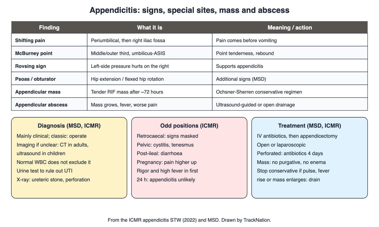
Appendicitis signs, special positions and treatment. Drawn by TrackNation.
Cause and Clinical Picture (MSD, ICMR)
Commonest surgical cause of acute abdominal pain; peaks in adolescence and the 20s. Luminal obstruction — usually lymphoid hyperplasia, sometimes a faecolith — → distension, bacterial overgrowth, ischaemia, gangrene, perforation; omentum walls it off as an abscess.
Classic: periumbilical/epigastric pain, then nausea/vomiting and anorexia; pain shifts to the right iliac fossa. ICMR: pain always precedes vomiting; fever usually absent in the first 6 hours; low-grade fever is common later.
Signs: pointing sign, cough pain in the RIF, McBurney point (junction of middle and outer thirds of umbilicus–ASIS line) tenderness, guarding, rebound, Rovsing (LIF pressure → RIF pain), psoas (pain on passive hip extension), obturator (pain on internal rotation of flexed thigh). Generalised rigidity = peritonitis.
Variants (ICMR): retrocaecal — signs masked; pelvic — cystitis or tenesmus; post-ileal — diarrhoea; pregnancy — pain higher as the caecum is pushed up; salpingitis mimics (dysmenorrhoea, discharge). Rigors and high fever within 24 h make appendicitis unlikely. Older people and pregnant women have atypical, milder signs.
Diagnosis (MSD, ICMR)
Diagnosis is clinical; with classic features, operate — imaging only delays and risks perforation.
Atypical cases: contrast CT (accurate, shows other causes) or graded-compression ultrasound (no radiation — preferred in children; also shows collections). Laparoscopy helps in women with unclear lower abdominal pain.
WBC usually raised but a normal count does not exclude appendicitis. Urinalysis to exclude UTI (RBC/WBC can appear in urine); plain X-ray to rule out ureteric stone or perforated ulcer (ICMR).
Higher complication risk: extremes of age, immunosuppression, diabetes, faecolith obstruction, previous surgery (ICMR).
Treatment, Mass and Abscess (ICMR, MSD)
IV fluids and antibiotics (third-generation cephalosporin) on admission, then appendicectomy — open or laparoscopic. Non-perforated: no further antibiotics; perforated: continue ~4 days. A 15% negative appendicectomy rate is acceptable because delay kills. Avoid appendicectomy if Crohn disease involves the caecum.
Antibiotics alone often settle the attack, but many recur within a year — appendicectomy is still advised, especially with an appendicolith. Early surgery mortality <1%.
Appendicular mass (late, ≥72 h): surgery risks bleeding, caecal/ileal injury and faecal fistula → Ochsner-Sherren conservative regimen: IV fluids, antibiotics, no purgatives, no enemas; improvement = less pain and tenderness, appetite back. Stop if pulse and temperature rise, tenderness spreads, the mass grows, or vomiting/large aspirates → ultrasound for pus → guided aspiration or open drainage. Operations for mass are done at a higher centre.
Abscess: percutaneous ultrasound-guided or open drainage, appendicectomy later. Large inflammatory mass found at surgery → resection with ileocolostomy. Post-op fever: check the wound, residual abscess (pelvis, RIF, Morison pouch), chest, thrombophlebitis, jaundice, UTI.
Recurrent appendicitis: repeated RIF pain, tender McBurney point; ultrasound/CT; appendicectomy. The appendix is also a common site of neuroendocrine tumours.
Traps the Examiner Sets
Vomiting before pain argues against appendicitis.
Normal WBC never rules it out.
Appendicular mass: conservative first — no purgatives or enemas.
Ultrasound for children; CT for unclear adults.
Memory Hooks & Shortcuts
Shortcuts to lock in this article's numbers and lists. Each one only re-packages facts proven above.
🧠 'Pain first, puke later' — reverse order, rethink — sequence
🧠 'Rovsing Reverses': press left, hurts right — signs
🧠 Positions: 'Retro hides, Pelvic pees, Post-ileal runs' — atypical
🧠 Ochsner-Sherren: 'Feed the drip, starve the gut, no enema, watch the pulse' — mass
🧠 'Kids get the probe (US), adults get the scanner (CT)' — imaging
Rapid-Revision Cards
Cause
Lymphoid hyperplasia, faecolith.
Sequence
Pain → vomiting; shifts to RIF.
McBurney
Middle/outer third umbilicus–ASIS.
Rovsing
LIF pressure → RIF pain.
Psoas sign
Hip extension hurts.
Obturator sign
Internal rotation of flexed thigh.
Retrocaecal
Signs masked; diarrhoea.
Pelvic
Cystitis, tenesmus.
Pregnancy
Pain higher.
Early rigors
Appendicitis unlikely.
Classic case
Operate; don't image.
Child imaging
Ultrasound.
Adult unclear
CT.
Normal WBC
Doesn't exclude.
Antibiotics
3rd-gen cephalosporin pre-op.
Perforated
Antibiotics 4 days.
Negative rate
15% acceptable.
Mass
Ochsner-Sherren; no enema.
Stop regimen
Pulse, fever up; mass grows.
Abscess
Drain; interval appendicectomy.
Crohn caecum
Leave the appendix.
Sources
Facts in this article were checked against these documents.
Standard Treatment Workflow: Appendicitis — ICMR / Department of Health Research, 2022
MSD Manual Professional: Appendicitis; Carcinoid Syndrome — Merck & Co. (MSD Manuals), 2025 (link)
Competency Based Undergraduate Curriculum, Vol III: General Surgery (SU28.15) — National Medical Commission, 2024
Test Yourself: Practice Questions
Thirty questions in NEET PG style, most of them clinical vignettes.
Practice Questions (NEET PG pattern)
Original questions written in the style of the exam: +4 for a correct answer, −1 for a wrong one. Every answer was checked against the sources listed above. NEET PG papers are not published by NBEMS, so these are not past-paper questions.
Q1. The commonest cause of acute abdominal pain requiring surgery is
(a) perforated ulcer
(b) acute appendicitis
(c) cholecystitis
(d) diverticulitis
Show answer
Answer: (b) — MSD.
Q2. Appendiceal luminal obstruction is most often due to
(a) worms
(b) lymphoid hyperplasia
(c) carcinoid
(d) foreign body
Show answer
Answer: (b) — Sometimes faecolith.
Q3. A teenager has periumbilical pain, then nausea and anorexia; after a few hours the pain moves to the right iliac fossa. This is
(a) mesenteric adenitis always
(b) renal colic
(c) gastroenteritis
(d) classic acute appendicitis
Show answer
Answer: (d) — Shifting pain.
Q4. According to the ICMR workflow, in acute appendicitis
(a) high fever starts the illness
(b) vomiting precedes pain
(c) pain always precedes vomiting
(d) diarrhoea is always present
Show answer
Answer: (c) — Murphy's sequence.
Q5. McBurney point lies at the
(a) tip of the 12th rib
(b) junction of the middle and outer thirds of the umbilicus–ASIS line
(c) midpoint of the inguinal ligament
(d) umbilicus
Show answer
Answer: (b) — Point tenderness.
Q6. Pain in the right iliac fossa on pressing the left iliac fossa is
(a) psoas sign
(b) obturator sign
(c) Rovsing sign
(d) Murphy sign
Show answer
Answer: (c) — Supports appendicitis.
Q7. Pain on passive extension of the right hip is the
(a) psoas sign
(b) obturator sign
(c) Kehr sign
(d) Rovsing sign
Show answer
Answer: (a) — Stretches iliopsoas.
Q8. Pain on passive internal rotation of the flexed right thigh is the
(a) psoas sign
(b) obturator sign
(c) Blumberg sign
(d) Cullen sign
Show answer
Answer: (b) — MSD.
Q9. In a retrocaecal appendicitis, the abdominal signs are
(a) only in the chest
(b) always florid
(c) on the left
(d) often masked
Show answer
Answer: (d) — ICMR.
Q10. A patient with appendicitis has frequency and dysuria with tenesmus. The appendix is most likely
(a) subhepatic
(b) retrocaecal
(c) pre-ileal
(d) pelvic
Show answer
Answer: (d) — Irritates bladder and rectum.
Q11. Diarrhoea with appendicitis suggests an appendix that is
(a) pelvic always
(b) normal
(c) subhepatic
(d) post-ileal or retrocaecal
Show answer
Answer: (d) — ICMR: post-ileal; MSD: retrocaecal.
Q12. In late pregnancy, appendicitis pain is often
(a) only in the back
(b) higher, towards the right flank
(c) absent
(d) in the left iliac fossa
Show answer
Answer: (b) — Caecum pushed up.
Q13. A patient has rigors and high fever within hours of the onset of abdominal pain. Per ICMR, appendicitis is
(a) certain
(b) most unlikely
(c) retrocaecal
(d) perforated
Show answer
Answer: (b) — Fever usually absent in first 6 h.
Q14. A young man has classic history and signs of appendicitis. The next step is
(a) CT first in every case
(b) MRI
(c) appendicectomy without delaying for imaging
(d) observe 48 hours
Show answer
Answer: (c) — Imaging delays raise perforation.
Q15. In a child with equivocal signs of appendicitis, the preferred first imaging is
(a) CT abdomen
(b) plain X-ray
(c) graded-compression ultrasound
(d) barium enema
Show answer
Answer: (c) — No radiation.
Q16. A woman with unclear lower abdominal pain and suspected appendicitis may benefit most from
(a) diagnostic laparoscopy
(b) barium meal
(c) colonoscopy
(d) watchful waiting at home
Show answer
Answer: (a) — Diagnosis and treatment.
Q17. A patient with typical signs has a normal white cell count. Correct interpretation:
(a) a normal WBC does not exclude appendicitis
(b) appendicitis is excluded
(c) it must be a perforation
(d) repeat in a week
Show answer
Answer: (a) — Highly variable.
Q18. Urinalysis in suspected appendicitis is mainly done to
(a) measure amylase
(b) diagnose appendicitis
(c) check for pregnancy only
(d) exclude urinary tract infection
Show answer
Answer: (d) — ICMR.
Q19. Per ICMR, a plain abdominal X-ray in suspected appendicitis helps to rule out
(a) faecolith only
(b) Meckel diverticulum
(c) pregnancy
(d) ureteric calculus or peptic perforation
Show answer
Answer: (d) — Differentials.
Q20. Before appendicectomy, MSD advises IV
(a) oral amoxicillin only
(b) vancomycin alone
(c) third-generation cephalosporin
(d) no antibiotics
Show answer
Answer: (c) — Preoperative.
Q21. After appendicectomy for a perforated appendix, antibiotics should continue for about
(a) 4 days
(b) 6 weeks
(c) none
(d) 1 day
Show answer
Answer: (a) — None for non-perforated.
Q22. A negative appendicectomy rate of about 15% is accepted because
(a) CT is never accurate
(b) the appendix is useless
(c) delayed treatment increases mortality
(d) it reduces cost
Show answer
Answer: (c) — MSD.
Q23. Treating uncomplicated appendicitis with antibiotics alone
(a) always cures permanently
(b) is contraindicated by law
(c) is preferred with an appendicolith
(d) often works initially but many recur within a year
Show answer
Answer: (d) — Appendicectomy still advised.
Q24. A patient presenting 5 days after onset has a tender RIF mass and is improving. Management is
(a) discharge without treatment
(b) enema and purgatives
(c) immediate appendicectomy
(d) Ochsner-Sherren conservative regimen
Show answer
Answer: (d) — Surgery risks injury and fistula.
Q25. During conservative treatment of an appendicular mass, which is avoided?
(a) IV fluids
(b) purgatives and enemas
(c) analgesia
(d) antibiotics
Show answer
Answer: (b) — ICMR.
Q26. Conservative treatment of an appendicular mass should be stopped if
(a) tenderness diminishes
(b) appetite returns
(c) pain lessens
(d) pulse and temperature rise and the mass enlarges
Show answer
Answer: (d) — Then drain.
Q27. An appendiceal abscess found late is best managed by
(a) enema
(b) oral antibiotics at home
(c) immediate right hemicolectomy
(d) ultrasound-guided percutaneous or open drainage, appendicectomy later
Show answer
Answer: (d) — Interval appendicectomy.
Q28. A patient has fever a week after appendicectomy. Per ICMR, a key site to look for a residual abscess is the
(a) thyroid
(b) left subphrenic space only
(c) pelvis, RIF or Morison pouch
(d) spleen
Show answer
Answer: (c) — Also wound, chest, UTI.
Q29. At surgery for appendicitis, the caecum is found involved by Crohn disease. MSD advises
(a) not removing the appendix
(b) removing the appendix always
(c) right hemicolectomy routinely
(d) leaving a drain only
Show answer
Answer: (a) — Fistula risk.
Q30. With early surgery in well-resourced settings, mortality from appendicitis is
(a) about 25%
(b) over 50%
(c) about 10%
(d) under 1%
Show answer
Answer: (d) — Worse with perforation, old age.
Active Recall
From memory: explain the cause and classic sequence of appendicitis, describe its signs and atypical positions, outline when and how to image, and describe antibiotic use, appendicectomy, the appendicular mass regimen and its stopping criteria, and abscess management.