Cell Injury, Adaptation and Cell Death for NEET PG Pathology: Apoptosis vs Necrosis, Necrosis Types, Calcification
Start Here
🎯 Exam priority: High. Expect: name the adaptation, apoptosis vs necrosis, caspases and the two apoptosis pathways, the newer death types (necroptosis, pyroptosis, ferroptosis), which necrosis goes with which cause, and dystrophic vs metastatic calcification.
CBME mapping: PA 1.1–1.3, 2.1–2.8. Cell-death terms follow the Nomenclature Committee on Cell Death (NCCD, 2018).
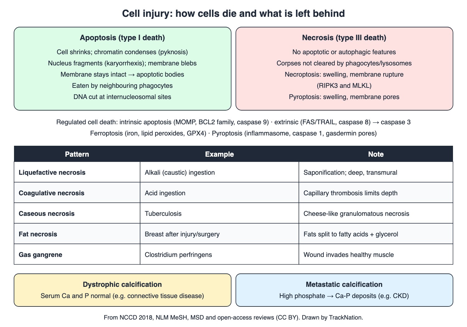
Apoptosis vs necrosis, regulated cell death, necrosis patterns and calcification. Drawn by TrackNation.
Adaptations
Adaptation (MeSH) | What changes |
Atrophy | Cell, tissue or organ shrinks (ischaemia, malnutrition, hormonal change) |
Hypertrophy | Bulk rises from bigger cells, not more cells |
Hyperplasia | More cells, without a tumour |
Metaplasia | One adult cell type replaced by another (e.g. Barrett oesophagus) |
Dysplasia (NCI) | Abnormal cells; not cancer but may become cancer |
Barrett oesophagus (squamous replaced by columnar epithelium after reflux) can progress to oesophageal adenocarcinoma.
Why Cells Get Hurt
Ischaemia (poor perfusion) and hypoxia (low oxygen). Free radicals such as reactive oxygen species have an unpaired electron and damage DNA, RNA and proteins. Reperfusion injury: harm when blood flow returns after ischaemia.
Cell cycle: G0 resting; G1 12 h to days; S (DNA synthesis) 2–4 h. Telomeres shorten with each division; senescence is permanent growth arrest without death, triggered by DNA damage or oxidative stress. Cancer cells use telomerase.
How Cells Die
Accidental cell death is instant destruction by severe physical, chemical or mechanical insults; regulated cell death uses dedicated machinery and can be modified by drugs or genes.
Apoptosis: shrinkage, chromatin condensation (pyknosis), nuclear fragmentation (karyorrhexis), membrane blebbing and apoptotic bodies that neighbouring cells eat. The membrane stays intact. DNA is cut at regular internucleosomal sites. It balances mitosis.
Intrinsic pathway: stress (DNA damage, growth-factor loss, ROS) → mitochondrial outer-membrane permeabilisation (MOMP), controlled by the BCL2 family → cytochrome c + APAF1 + caspase 9 form the apoptosome. Extrinsic pathway: death receptors (FAS with FAS ligand, TRAIL receptors) → caspase 8. Both end in executioner caspase 3, which exposes phosphatidylserine. Caspases are cysteine proteases that cut after aspartate.
Necrosis (classical type III): no apoptotic features, and the debris is not cleared by phagocytes. Necroptosis: RIPK3 and MLKL; swelling and membrane rupture. Pyroptosis: inflammasome, caspase 1, gasdermin pores, inflammation. Ferroptosis: iron-dependent lipid peroxidation, controlled by GPX4. Dying cells release danger signals (DAMPs). Autolysis: tissue breakdown by its own enzymes.
Patterns of Necrosis and Gangrene
Liquefactive: alkali ingestion — saponification, deep transmural damage. Coagulative: acid ingestion — proteins denature and capillaries thrombose, which limits penetration.
Caseous: tuberculosis — cheese-like granulomatous necrosis with epithelioid granulomas.
Fat necrosis: fats split into fatty acids and glycerol; in the breast after injury, surgery or radiation it can form a firm, round lump.
Gangrene: death and putrefaction of tissue from lost blood supply. Gas gangrene: Clostridium (C. perfringens in over 80%) spreading from a contaminated wound into healthy muscle. Infarction: necrosis from local ischaemia, usually thrombus or embolus.
Accumulations, Pigments and Calcification
Fatty liver: triglyceride droplets make the liver yellow. Lipofuscin: lipid pigment that builds up with age. Haemosiderosis: excess stainable iron in liver and macrophages without tissue damage. Hyalin: clear, homogeneous, eosinophilic material. Melanin protects skin from UV.
Calcification: dystrophic occurs with normal serum calcium and phosphate (the commonest type in connective tissue disease); metastatic comes from high phosphate depositing calcium phosphate, as in chronic kidney disease. Vessel calcification accompanies atherosclerotic plaques. Psammoma bodies are concentric calcified rings in some tumours.
Traps the Examiner Sets
Hypertrophy = bigger cells; hyperplasia = more cells.
Alkali = liquefactive (deeper); acid = coagulative (shallower).
Apoptosis keeps the membrane intact; necroptosis and pyroptosis rupture it.
Intrinsic = caspase 9 (mitochondria); extrinsic = caspase 8 (FAS); both → caspase 3.
Dystrophic = normal calcium; metastatic = deranged calcium–phosphate.
Memory Hooks & Shortcuts
Shortcuts to lock in this article's numbers and lists. Each one only re-packages facts proven above.
🧠 Hyper-TROPHY = Trophy-sized cells; Hyper-PLASIA = Plenty of cells — hypertrophy vs hyperplasia
🧠 Apoptosis is a tidy suicide: Shrink, Shatter, get Swallowed — apoptosis morphology
🧠 Nine from the mitochondria, Eight from the doorbell (FAS), Three finishes the job — caspases 9, 8, 3
🧠 Pyro = fire = Inflammasome and caspase 1 — pyroptosis
🧠 Ferro = iron = rusting lipids — ferroptosis
🧠 ALKali = ALL the way through (liquefactive) — alkali vs acid necrosis
🧠 Dystrophic = Damaged tissue, normal blood; Metastatic = Minerals high — calcification
Rapid-Revision Cards
Atrophy
Shrinkage — ischaemia, malnutrition, hormones.
Hypertrophy vs hyperplasia
Bigger cells vs more cells.
Metaplasia
Adult cell type swapped — Barrett.
Dysplasia
Abnormal cells, not yet cancer.
Reperfusion injury
Damage when flow returns after ischaemia.
Free radicals
Unpaired electron; damage DNA, proteins.
S phase
DNA synthesis, 2–4 h.
Senescence
Permanent arrest, cell alive.
Apoptosis morphology
Shrinkage, pyknosis, karyorrhexis, blebs, apoptotic bodies.
Apoptosis membrane
Intact; cleared by phagocytes.
Intrinsic pathway
MOMP (BCL2) → cytochrome c + APAF1 → caspase 9.
Extrinsic pathway
FAS/TRAIL → caspase 8.
Executioner
Caspase 3; exposes phosphatidylserine.
Necroptosis
RIPK3, MLKL; rupture.
Pyroptosis
Inflammasome, caspase 1, gasdermin.
Ferroptosis
Iron, lipid peroxides, GPX4.
Alkali vs acid
Liquefactive deep vs coagulative shallow.
Caseous
TB; cheese-like.
Fat necrosis
Fatty acids + glycerol; breast lump.
Gas gangrene
C. perfringens > 80%.
Lipofuscin
Ageing pigment.
Haemosiderosis
Iron, no damage.
Dystrophic vs metastatic
Normal Ca/P vs high phosphate.
Psammoma bodies
Concentric calcified rings.
Sources
Facts in this article were checked against these documents.
Molecular mechanisms of cell death: recommendations of the Nomenclature Committee on Cell Death 2018 — Galluzzi L et al., Cell Death Differ 25:486–541, 2018 (link)
Medical Subject Headings (MeSH) scope notes — US National Library of Medicine, accessed 2026 (link)
NCI Dictionary of Cancer Terms — National Cancer Institute, accessed 2026 (link)
MSD Manual Professional: Tuberculosis; GERD; Hyperphosphatemia; Cellular and Molecular Basis of Cancer — Merck & Co. (MSD Manuals), reviewed 2025–26 (link)
Post-Caustic Esophagitis: Updated Clinical Insights (J Clin Med, CC BY) — Europe PMC PMC12693313, 2025 (link)
Calcinosis Cutis Universalis: A Review (CC BY); Phosphate: An Anion Controlling Metabolic Functions (CC BY) — Europe PMC PMC12897785; PMC13467933, 2026 (link)
Competency Based Undergraduate Curriculum, Pathology (PA 1–2) — National Medical Commission, 2024
Test Yourself: Practice Questions
Thirty-three questions in NEET PG style, most of them clinical vignettes.
Practice Questions (NEET PG pattern)
Original questions written in the style of the exam: +4 for a correct answer, −1 for a wrong one. Every answer was checked against the sources listed above. NEET PG papers are not published by NBEMS, so these are not past-paper questions.
Q1. A tissue shrinks because of long-standing malnutrition and poor blood supply. This change is
(a) metaplasia
(b) atrophy
(c) hyperplasia
(d) hypertrophy
Show answer
Answer: (b) — Decrease in size of a cell, tissue or organ.
Q2. An organ enlarges because its cells grow bigger, without any increase in cell number. This is
(a) dysplasia
(b) hyperplasia
(c) hypertrophy
(d) metaplasia
Show answer
Answer: (c) — Hyperplasia = more cells.
Q3. An increase in the number of normal-looking cells in a tissue, without a tumour, is
(a) atrophy
(b) hyperplasia
(c) anaplasia
(d) hypertrophy
Show answer
Answer: (b) — MeSH.
Q4. A man with long-standing acid reflux has columnar epithelium replacing the squamous lining of the distal oesophagus. This is
(a) hyperplasia only
(b) dysplasia
(c) normal ageing
(d) metaplasia (Barrett oesophagus), which can lead to adenocarcinoma
Show answer
Answer: (d) — One adult cell type replaced by another.
Q5. A biopsy shows abnormal cells that are not cancer but may become cancer. The term is
(a) atrophy
(b) metaplasia
(c) dysplasia
(d) hyperplasia
Show answer
Answer: (c) — Graded mild, moderate or severe.
Q6. Blood flow returns to a tissue after a period of ischaemia, and further damage follows. This is
(a) apoptosis only
(b) reperfusion injury
(c) autolysis
(d) atrophy
Show answer
Answer: (b) — Restoration of blood flow after ischaemia.
Q7. Reactive oxygen species damage cells because they
(a) are enzymes that cut DNA
(b) block caspases
(c) are free radicals with an unpaired electron
(d) are antibodies
Show answer
Answer: (c) — They damage DNA, RNA and proteins.
Q8. In the cell cycle, DNA is synthesised during
(a) G1
(b) S phase (usually 2–4 h)
(c) M phase
(d) G0
Show answer
Answer: (b) — G1 lasts 12 h to a few days.
Q9. Cells that have permanently stopped dividing but are still alive, after DNA damage or oxidative stress, are in
(a) mitosis
(b) necrosis
(c) apoptosis
(d) senescence
Show answer
Answer: (d) — Telomeres shorten with each division.
Q10. A cell shrinks, its chromatin condenses, the nucleus fragments and small membrane-bound bodies are eaten by neighbouring cells. This is
(a) necrosis
(b) autolysis
(c) apoptosis
(d) liquefaction
Show answer
Answer: (c) — Pyknosis, karyorrhexis, apoptotic bodies; membrane stays intact.
Q11. A feature that distinguishes apoptosis from necrosis is that in apoptosis the
(a) lysosomes are not involved
(b) debris is never removed
(c) plasma membrane stays intact and the cell is cleared by phagocytes
(d) cell swells and bursts
Show answer
Answer: (c) — Classical necrosis has no phagocytic clearance.
Q12. In apoptosis, genomic DNA is cut
(a) at regularly spaced internucleosomal sites
(b) only at telomeres
(c) randomly
(d) by lysosomal enzymes after death
Show answer
Answer: (a) — MeSH definition of apoptosis.
Q13. In the intrinsic (mitochondrial) pathway of apoptosis, the committing step is
(a) mitochondrial outer membrane permeabilisation, controlled by the BCL2 family
(b) activation of caspase 1
(c) lipid peroxidation
(d) binding of FAS ligand
Show answer
Answer: (a) — Cytochrome c is then released.
Q14. Cytochrome c released from mitochondria joins APAF1 to form the apoptosome, which activates
(a) RIPK3
(b) caspase 8
(c) caspase 1
(d) caspase 9
Show answer
Answer: (d) — Initiator of the intrinsic pathway.
Q15. A cell dies after FAS ligand binds its FAS receptor. The initiator caspase is
(a) caspase 9
(b) caspase 1
(c) caspase 8 (extrinsic pathway)
(d) caspase 3 alone
Show answer
Answer: (c) — Both pathways end in executioner caspase 3.
Q16. Caspases are
(a) lipases
(b) serine proteases
(c) DNA polymerases
(d) cysteine proteases that cut after an aspartic acid residue
Show answer
Answer: (d) — Activated by cleavage of precursors.
Q17. Regulated cell death that depends on RIPK3 and MLKL and ends in cell swelling and membrane rupture is
(a) necroptosis
(b) autophagy
(c) ferroptosis
(d) apoptosis
Show answer
Answer: (a) — Can be triggered via death receptors such as TNFR1.
Q18. A cell infected by an intracellular pathogen dies with inflammasome formation, caspase 1 activation and cytokine release. This is
(a) pyroptosis
(b) necroptosis
(c) apoptosis
(d) ferroptosis
Show answer
Answer: (a) — Gasdermin forms membrane pores.
Q19. Cell death driven by iron-dependent lipid peroxidation and blocked by iron chelators is
(a) autolysis
(b) ferroptosis (controlled by GPX4)
(c) apoptosis
(d) pyroptosis
Show answer
Answer: (b) — Lipophilic antioxidants also inhibit it.
Q20. Breakdown of tissue after death by the cells' own enzymes is called
(a) autolysis
(b) apoptosis
(c) caseation
(d) heterolysis by bacteria
Show answer
Answer: (a) — MeSH.
Q21. A child swallows a strong alkali. The type of necrosis expected in the oesophagus is
(a) coagulative necrosis limited to the surface
(b) fat necrosis
(c) caseous necrosis
(d) liquefactive necrosis with deep, transmural injury
Show answer
Answer: (d) — Alkalis saponify fats and proteins.
Q22. Strong acid ingestion produces which type of necrosis, and why is it shallower?
(a) liquefactive — saponification
(b) coagulative necrosis — capillary thrombosis limits penetration
(c) fibrinoid — immune complexes
(d) caseous — granulomas
Show answer
Answer: (b) — Acids convert tissue proteins and thrombose small vessels.
Q23. A lymph node biopsy shows epithelioid granulomas with cheese-like necrotic centres. This necrosis is typical of
(a) acid burns
(b) gas gangrene
(c) acute pancreatitis
(d) tuberculosis (caseous necrosis)
Show answer
Answer: (d) — Delayed hypersensitivity response.
Q24. A woman notices a firm breast lump months after breast surgery; biopsy shows dead fat tissue. This is
(a) fat necrosis
(b) caseous necrosis
(c) gangrene
(d) liquefactive necrosis
Show answer
Answer: (a) — Fats split into fatty acids and glycerol.
Q25. A contaminated wound develops infection spreading into healthy muscle. The organism in most cases is
(a) Streptococcus pneumoniae
(b) Mycobacterium tuberculosis
(c) Staphylococcus aureus
(d) Clostridium perfringens (gas gangrene)
Show answer
Answer: (d) — Over 80% of cases.
Q26. Death and putrefaction of tissue due to loss of blood supply is
(a) autolysis
(b) gangrene
(c) atrophy
(d) metaplasia
Show answer
Answer: (b) — Gas gangrene is a clostridial infection.
Q27. An infarct is
(a) fat accumulation
(b) apoptosis from hormone loss
(c) necrosis from local ischaemia, usually due to a thrombus or embolus
(d) calcification of normal tissue
Show answer
Answer: (c) — MeSH.
Q28. A lipid pigment that accumulates in normal tissues and increases with age is
(a) haemosiderin
(b) bilirubin
(c) lipofuscin
(d) melanin
Show answer
Answer: (c) — MeSH: similar to ceroid.
Q29. A patient has increased stainable iron in the liver and macrophages without organ damage. This is
(a) lipofuscinosis
(b) fatty liver
(c) haemosiderosis
(d) haemochromatosis with cirrhosis
Show answer
Answer: (c) — Iron is stored as haemosiderin.
Q30. A patient with a connective tissue disease has calcium deposits in the skin with normal serum calcium and phosphate. The type is
(a) metastatic calcification
(b) dystrophic calcification
(c) ossification
(d) calciphylaxis only
Show answer
Answer: (b) — The commonest subtype in connective tissue disease.
Q31. A dialysis patient with persistently high phosphate develops hard calcium deposits in soft tissues. This is
(a) dystrophic calcification
(b) psammoma body formation
(c) metastatic calcification
(d) fat necrosis
Show answer
Answer: (c) — Calcium-phosphate deposits in connective tissue.
Q32. Microscopy of a tumour shows small laminated calcified bodies with concentric rings. These are
(a) Heinz bodies
(b) Russell bodies
(c) psammoma bodies
(d) apoptotic bodies
Show answer
Answer: (c) — Seen in some benign and malignant tumours.
Q33. A liver biopsy shows hepatocytes filled with lipid droplets, single large or many small; the liver looks yellow. The lipid is mainly
(a) cholesterol esters only
(b) lipofuscin
(c) triglyceride (fatty liver)
(d) glycogen
Show answer
Answer: (c) — Caused by an imbalance in fatty-acid metabolism.
Active Recall
From memory: define the four adaptations with an example of metaplasia; explain reperfusion injury and senescence; describe apoptosis morphology and the intrinsic and extrinsic pathways with their caspases; contrast necroptosis, pyroptosis and ferroptosis; match each necrosis pattern to a cause; and distinguish dystrophic from metastatic calcification.