Haemodynamic Disorders for NEET PG Pathology: Oedema, Shock, Virchow Triad, Thrombophilia, Embolism, Infarction
Start Here
🎯 Exam priority: High. Expect: mechanism of oedema in a scenario, the type of shock, Virchow triad, factor V Leiden, the source of pulmonary emboli, paradoxical and amniotic fluid embolism, and why lung infarcts are uncommon.
CBME mapping: PA 5.1–5.6. Normal haemostasis and bleeding disorders are in article 14.
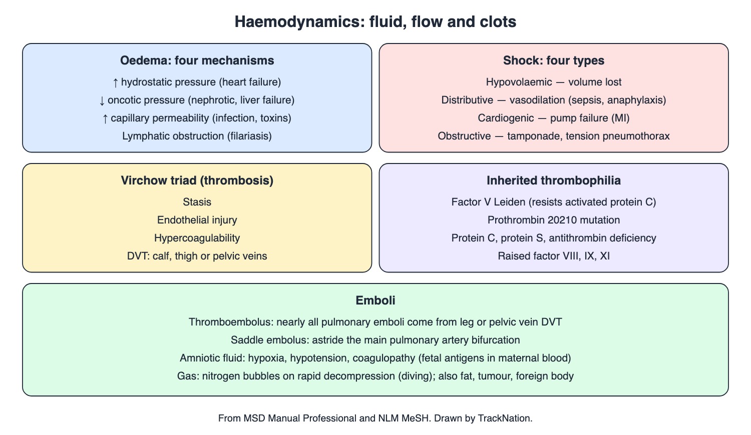
Oedema, shock, Virchow triad, thrombophilia and emboli. Drawn by TrackNation.
Oedema
Oedema is extra interstitial fluid — mostly water, but protein- and cell-rich with infection or lymphatic block. Four mechanisms: raised hydrostatic pressure (heart failure: symmetric, dependent, pitting); low oncotic pressure (nephrotic syndrome, protein-losing enteropathy, liver failure, starvation); raised permeability (infection, toxins, inflammation); lymphatic obstruction (lymphoedema; filariasis in endemic areas, sometimes genital).
Ascites = free fluid in the peritoneal cavity. Pulmonary oedema impairs gas exchange. Cytotoxic brain oedema (intracellular) follows hypoxic or ischaemic injury.
Hyperaemia, Congestion and Haemorrhage
Hyperaemia (active, arterial) = more inflow; congestion (passive, venous) = blocked outflow.
Haemorrhage = escape of blood from a vessel. Petechiae (pinpoint) < purpura < ecchymosis (> 2–3 cm, flat bruise); a haematoma is a collection of blood outside vessels.
Shock
Shock is failure to perfuse or oxygenate vital organs: cool clammy skin, confusion, lactate > 2 mmol/L, urine < 0.5 mL/kg/h. Hypotension (SBP < 90 or MAP < 65) is common but not required. Cells switch to anaerobic metabolism; if shock persists, damage becomes irreversible. In septic (distributive) shock the skin may be warm and flushed; septic shock is sepsis with hypotension or hypoperfusion despite fluids.
Shock type (MSD) | Mechanism | Examples |
Hypovolaemic | Critical loss of intravascular volume | Haemorrhage, fluid loss |
Distributive | Vasodilation; blood volume normal | Sepsis, anaphylaxis, toxic shock, spinal injury above T4 |
Cardiogenic | Primary cardiac failure of output | Myocardial infarction, arrhythmia |
Obstructive | Mechanical block to filling or emptying | Tension pneumothorax, cardiac tamponade |
Thrombosis
Virchow triad: stasis, endothelial injury, hypercoagulability. DVT begins in venous valve cusps; venous thrombi are red (fibrin and red cells, few platelets). Causes: immobility, leg fractures, cancer.
Inherited thrombophilia: factor V Leiden (R506Q; factor Va resists activated protein C — the commonest cause of APC resistance), prothrombin 20210 mutation, deficiency of protein C, protein S or antithrombin, raised factors VIII, IX and XI.
Embolism and Infarction
Embolus (MSD/MeSH) | Key fact |
Thromboembolus (PE) | Nearly all from leg or pelvic vein DVT; higher risk at popliteal level or above |
Saddle | Lodges at the bifurcation of the main pulmonary artery |
Paradoxical | Venous clot crosses to the arterial side via ASD or patent foramen ovale → stroke |
Fat | After fractures of large bones, or corticosteroids |
Air | After trauma, surgery or pressure change |
Amniotic fluid | Hypoxia, hypotension, coagulopathy; a leading cause of sudden death in labour |
Decompression (caisson disease, the bends) | Nitrogen bubbles on rapid ascent; joints and circulation; recompression |
Infarction is necrosis from local ischaemia, usually a thrombus or embolus. Pulmonary infarcts appear as pleural-based wedges but are uncommon because the lung has a dual (bronchial and pulmonary) supply. A negative D-dimer makes PE very unlikely when pre-test probability is low.
Traps the Examiner Sets
Nephrotic or liver failure oedema = low oncotic pressure; heart failure = high hydrostatic pressure.
Septic shock is distributive (warm skin early); tamponade is obstructive.
Pulmonary emboli come from leg/pelvic veins; paradoxical emboli need a right-to-left shunt.
Lung infarcts are rare: dual blood supply.
Memory Hooks & Shortcuts
Shortcuts to lock in this article's numbers and lists. Each one only re-packages facts proven above.
🧠 Pump, Pipe, Plasma, Plumbing — hydrostatic, permeability, oncotic, lymphatic oedema
🧠 HDCO: Hole (volume lost), Dilated, Can't pump, Obstructed — four shock types
🧠 Virchow's SEH: Stasis, Endothelium, Hypercoagulable — Virchow triad
🧠 Leiden Leaves Va alone (APC can't cut it) — factor V Leiden
🧠 Paradox needs a Passage (PFO/ASD) — paradoxical embolism
🧠 FAT after Femur, AIR after Ascent — fat and gas emboli
🧠 Two pipes save the lung — dual blood supply
Rapid-Revision Cards
Oedema mechanisms
↑ hydrostatic, ↓ oncotic, ↑ permeability, lymphatic block.
Nephrotic/liver oedema
Low oncotic pressure.
Heart-failure oedema
Symmetric, dependent, pitting — high hydrostatic.
Filariasis
Lymphoedema, sometimes genital.
Cytotoxic brain oedema
Intracellular; hypoxia/ischaemia.
Hyperaemia vs congestion
Active inflow vs passive outflow block.
Ecchymosis
> 2–3 cm flat bleed.
Shock types
Hypovolaemic, distributive, cardiogenic, obstructive.
Distributive
Vasodilation, normal volume — sepsis, anaphylaxis, neurogenic.
Obstructive
Tamponade, tension pneumothorax.
Shock markers
Lactate > 2; urine < 0.5 mL/kg/h; SBP < 90 or MAP < 65.
Virchow triad
Stasis, endothelial injury, hypercoagulability.
Venous thrombus
Red; valve cusps; few platelets.
Factor V Leiden
R506Q; APC resistance.
PE source
Leg/pelvic DVT.
Saddle embolus
Pulmonary artery bifurcation.
Paradoxical embolus
ASD/PFO → stroke.
Fat embolus
Large-bone fractures.
Amniotic fluid embolism
Hypoxia, hypotension, coagulopathy in labour.
Decompression sickness
Nitrogen bubbles; recompression.
Lung infarcts rare
Dual blood supply.
Sources
Facts in this article were checked against these documents.
MSD Manual Professional: Edema; Shock; Deep Venous Thrombosis; Pulmonary Embolism — Merck & Co. (MSD Manuals), reviewed 2025–26 (link)
MSD Manual Professional: Amniotic Fluid Embolism; Decompression Sickness; Overview of Thrombotic Disorders — Merck & Co. (MSD Manuals), reviewed 2025–26 (link)
Medical Subject Headings (MeSH) scope notes — US National Library of Medicine, accessed 2026 (link)
Competency Based Undergraduate Curriculum, Pathology (PA 5) — National Medical Commission, 2024
Test Yourself: Practice Questions
Thirty questions in NEET PG style, most of them clinical vignettes.
Practice Questions (NEET PG pattern)
Original questions written in the style of the exam: +4 for a correct answer, −1 for a wrong one. Every answer was checked against the sources listed above. NEET PG papers are not published by NBEMS, so these are not past-paper questions.
Q1. A child with nephrotic syndrome has generalised oedema. The main mechanism is
(a) increased capillary permeability from infection
(b) low plasma oncotic pressure
(c) lymphatic obstruction
(d) raised capillary hydrostatic pressure
Show answer
Answer: (b) — Also liver failure, protein-losing enteropathy, starvation.
Q2. A man with right heart failure has symmetric, dependent, painless, pitting leg oedema. The mechanism is
(a) raised capillary hydrostatic (venous) pressure
(b) lymphatic obstruction
(c) allergic permeability
(d) low oncotic pressure
Show answer
Answer: (a) — MSD oedema table.
Q3. A man from a filaria-endemic area has long-standing swelling of one leg and the scrotum. The mechanism is
(a) venous thrombosis
(b) low oncotic pressure
(c) right heart failure
(d) lymphatic obstruction (lymphoedema)
Show answer
Answer: (d) — Initially pitting, later fibrotic.
Q4. Oedema fluid that is rich in protein and cells points to
(a) nephrotic syndrome
(b) infection or lymphatic obstruction
(c) starvation
(d) heart failure
Show answer
Answer: (b) — Ordinary oedema fluid is mostly water.
Q5. After a severe hypoxic-ischaemic insult, the brain swells from fluid inside the cells. This is
(a) vasogenic oedema only
(b) cytotoxic brain oedema from hypoxic-ischaemic injury
(c) lymphoedema
(d) hydrocephalus
Show answer
Answer: (b) — MeSH.
Q6. Increased blood in an organ because venous outflow is obstructed is called
(a) active hyperaemia
(b) passive (venous) congestion
(c) infarction
(d) haemorrhage
Show answer
Answer: (b) — Active hyperaemia = more arterial inflow.
Q7. A flat purplish skin bleed larger than 2–3 cm is called
(a) a petechia
(b) purpura simplex only
(c) an ecchymosis
(d) a haematoma
Show answer
Answer: (c) — Larger than petechiae and purpura.
Q8. A collection of blood outside blood vessels, within a tissue or space, is a
(a) embolus
(b) petechia
(c) thrombus
(d) haematoma
Show answer
Answer: (d) — MeSH.
Q9. A patient with pneumonia has hypotension despite adequate fluids, warm flushed skin and a bounding pulse. The shock type is
(a) hypovolaemic
(b) obstructive
(c) distributive (septic)
(d) cardiogenic
Show answer
Answer: (c) — Vasodilation with normal blood volume.
Q10. Distributive shock differs from hypovolaemic shock because
(a) the heart fails as a pump
(b) blood volume is lost
(c) blood volume is normal but vessels are dilated
(d) great vessels are blocked
Show answer
Answer: (c) — Sepsis, anaphylaxis, toxic shock, spinal injury above T4.
Q11. After a large myocardial infarction, a patient develops shock from failure of cardiac output. The shock type is
(a) obstructive
(b) cardiogenic
(c) distributive
(d) hypovolaemic
Show answer
Answer: (b) — Primary cardiac failure of output.
Q12. A trauma patient is in shock caused by cardiac tamponade. The shock type is
(a) hypovolaemic
(b) obstructive
(c) cardiogenic
(d) distributive
Show answer
Answer: (b) — Also tension pneumothorax.
Q13. A patient with a high spinal cord injury (above T4) becomes hypotensive. The shock is
(a) obstructive
(b) cardiogenic
(c) neurogenic — a form of distributive shock
(d) hypovolaemic always
Show answer
Answer: (c) — MSD classifies it as distributive.
Q14. A laboratory marker of tissue hypoperfusion in shock is a blood lactate above
(a) 10 mmol/L
(b) 20 mmol/L
(c) 0.5 mmol/L
(d) 2.0 mmol/L
Show answer
Answer: (d) — Also oliguria < 0.5 mL/kg/h.
Q15. Hypotension in shock is usually defined as
(a) systolic < 90 mm Hg or mean arterial pressure < 65 mm Hg
(b) systolic < 120
(c) diastolic < 80
(d) mean arterial pressure < 100
Show answer
Answer: (a) — Common but not required for shock.
Q16. Septic shock is defined as sepsis with
(a) hypotension or hypoperfusion despite adequate fluid resuscitation
(b) a raised white count
(c) fever alone
(d) bacteraemia alone
Show answer
Answer: (a) — Lactic acidosis, oliguria, confusion.
Q17. The three factors of Virchow's triad are
(a) fever, pain and swelling
(b) anaemia, jaundice and splenomegaly
(c) stasis, endothelial injury and hypercoagulability
(d) hypertension, diabetes and smoking
Show answer
Answer: (c) — MSD DVT page.
Q18. An immobilised patient after a leg fracture develops a calf DVT. The thrombus is typically
(a) red — fibrin and red cells with few platelets, starting at valve cusps
(b) made of fat
(c) infected
(d) white — platelets only
Show answer
Answer: (a) — Immobility and leg fracture are classic causes.
Q19. A young woman with recurrent DVT has a mutation that makes activated factor V resistant to activated protein C. This is
(a) prothrombin 20210 mutation
(b) haemophilia
(c) factor V Leiden (R506Q)
(d) antithrombin deficiency
Show answer
Answer: (c) — The commonest cause of APC resistance.
Q20. Which of these is an inherited cause of venous thrombosis listed by MSD?
(a) protein C or protein S deficiency
(b) vitamin K deficiency
(c) haemophilia A
(d) von Willebrand disease
Show answer
Answer: (a) — Also antithrombin deficiency and prothrombin 20210.
Q21. Nearly all pulmonary emboli arise from
(a) the portal vein
(b) the left atrium
(c) deep vein thrombi in the legs or pelvis
(d) arm veins
Show answer
Answer: (c) — Risk higher if the thrombus reaches the popliteal vein or above.
Q22. A large embolus is found straddling the bifurcation of the main pulmonary artery. This is a
(a) fat embolus
(b) paradoxical embolus
(c) saddle embolus
(d) air embolus
Show answer
Answer: (c) — MSD.
Q23. A man with a leg DVT suddenly has a stroke. Echocardiography shows a patent foramen ovale. This is
(a) paradoxical embolism
(b) fat embolism
(c) septic embolism
(d) air embolism
Show answer
Answer: (a) — Venous clot reaches arterial side through a right-to-left defect.
Q24. After a fracture of a large bone, a patient develops an embolus that is not a blood clot. The classic type is
(a) air embolism
(b) tumour embolism
(c) amniotic fluid embolism
(d) fat embolism
Show answer
Answer: (d) — Also after corticosteroids (MeSH).
Q25. During labour, a woman suddenly develops hypoxia, hypotension and bleeding from a coagulopathy. The likely cause is
(a) amniotic fluid embolism
(b) pulmonary thromboembolism
(c) placenta praevia
(d) eclampsia
Show answer
Answer: (a) — Fetal antigens enter maternal blood; a leading cause of sudden death in labour.
Q26. A diver who surfaced rapidly develops severe joint pain and confusion. The cause and treatment are
(a) fat embolism — steroids
(b) air in the pleura — chest drain
(c) hypoglycaemia — glucose
(d) nitrogen bubbles from rapid decompression — recompression therapy
Show answer
Answer: (d) — Decompression sickness (caisson disease, the bends).
Q27. Air embolism usually follows
(a) large-bone fractures
(b) labour
(c) DVT
(d) trauma, surgery or changes in atmospheric pressure
Show answer
Answer: (d) — MeSH.
Q28. Pulmonary infarction is uncommon after pulmonary embolism because
(a) the lung has no veins
(b) emboli always dissolve
(c) the lung has a dual blood supply (bronchial and pulmonary)
(d) lung tissue does not need oxygen
Show answer
Answer: (c) — When it happens, it is a pleural-based wedge.
Q29. An infarct is
(a) fluid in tissues
(b) a collection of blood outside vessels
(c) necrosis from local ischaemia, usually due to a thrombus or embolus
(d) a type of shock
Show answer
Answer: (c) — MeSH.
Q30. In an outpatient with low pre-test probability of PE, a negative D-dimer
(a) means DVT is present
(b) makes pulmonary embolism very unlikely
(c) is useless
(d) confirms PE
Show answer
Answer: (b) — Below 0.4 mcg/mL.
Active Recall
From memory: give the four mechanisms of oedema with an example each; distinguish hyperaemia and congestion; classify the four types of shock with causes and markers; state Virchow triad and the inherited thrombophilias; and describe pulmonary, saddle, paradoxical, fat, air and amniotic fluid emboli, decompression sickness and why lung infarcts are uncommon.