Skin Basics for NEET PG Dermatology: Primary and Secondary Lesions, Bedside Tests and Topical Steroids
Start Here
🎯 Exam priority: Medium (foundation). Expect: size cut-offs for macule/papule/plaque and vesicle/bulla, which lesions blanch, palpable purpura, lichenification, keloid vs scar, Koebner phenomenon, KOH and Tzanck findings, Wood light colours, diascopy and ICMR topical steroid rules.
Every later Dermatology article uses these words. Skin histology is in Anatomy; inflammation in Pathology.
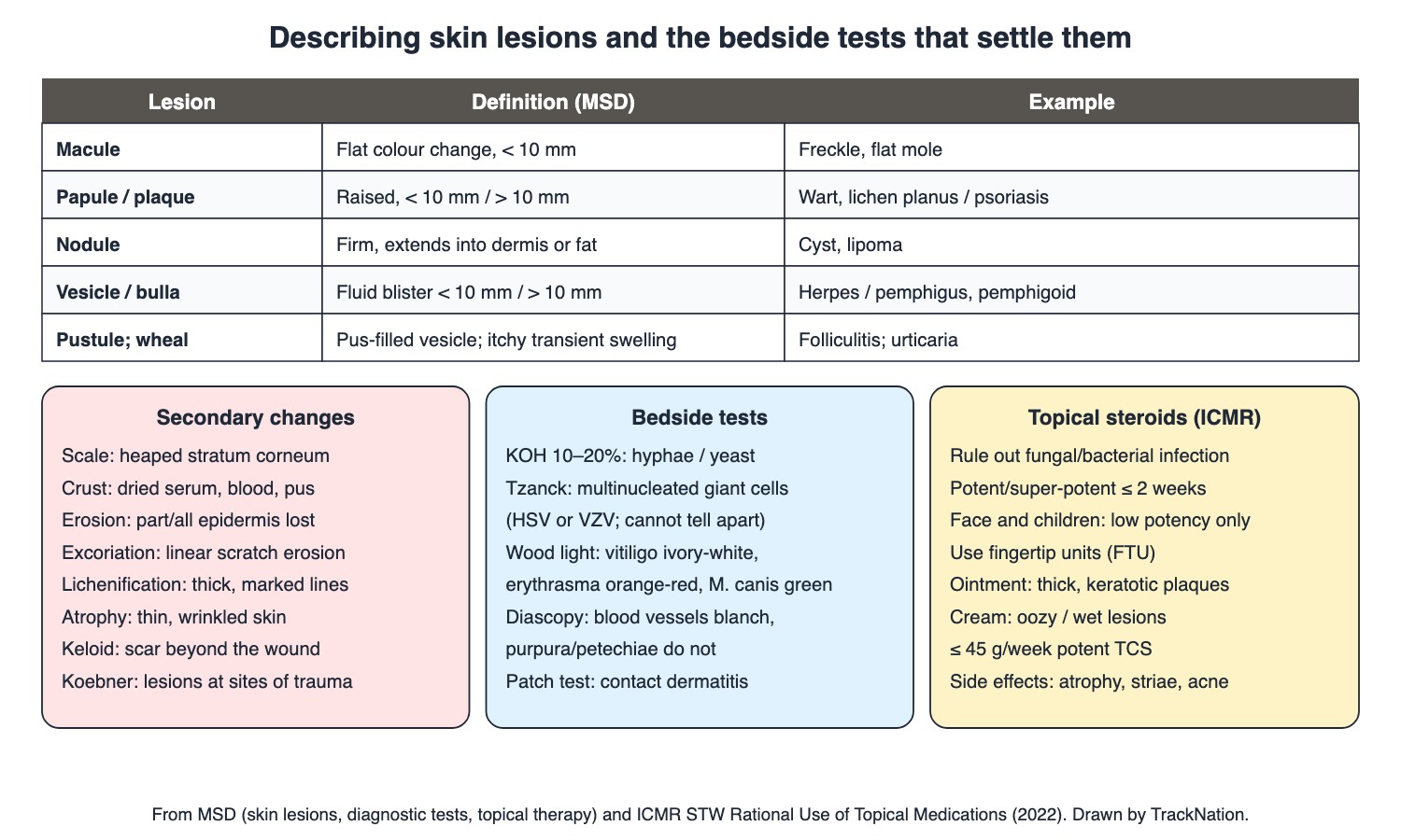
Skin lesions, bedside tests and topical steroid rules. Drawn by TrackNation.
Primary and Secondary Lesions
Macule: flat, non-palpable colour change < 10 mm (freckle, flat mole). Papule: raised, palpable < 10 mm. Plaque: palpable > 10 mm (psoriasis). Nodule: firm lesion extending into dermis or subcutis (cyst, lipoma).
Vesicle: fluid blister < 10 mm (herpes, acute contact dermatitis); bulla: > 10 mm (pemphigus vulgaris, bullous pemphigoid, burns, drug reactions). Pustule: vesicle with pus. Wheal: itchy, red, transient swelling.
Vascular: telangiectasia (permanently dilated small vessels); petechiae and purpura do not blanch; palpable purpura is the hallmark of leukocytoclastic vasculitis.
Secondary: scale (heaped stratum corneum), crust (dried serum, blood, pus), erosion (epidermal loss), excoriation (linear scratch), lichenification (thickening from rubbing), atrophy, scar; keloid extends beyond the wound margin. Annular = ring with central clearing.
Koebner phenomenon: new lesions at sites of trauma — psoriasis, lichen planus.
Bedside Tests and Topical Steroids
Test (MSD) | What it shows |
KOH mount (10–20%) | Hyphae or budding yeast → tinea or candidiasis |
Scabies scraping | Oil over a burrow or papule, unroof with a scalpel, look for mites |
Tzanck smear | Multinucleated giant cells → herpes simplex or zoster (cannot tell which) |
Wood light | Vitiligo ivory-white; erythrasma orange-red; M. canis tinea capitis green; Pseudomonas green; Trichophyton does not glow |
Diascopy | Vessels blanch; petechiae/purpura do not; sarcoid looks 'apple jelly' |
Patch test | Contact dermatitis allergen |
Punch biopsy | Usually 4 mm core into deep dermis/fat |
ICMR topical steroid rules: rule out local fungal or bacterial infection; potent and super-potent steroids for at most 2 weeks; only low potency on the face and in children; dose by fingertip units — whole adult body 20–30 g per application, one hand 0.3 g; no more than 45 g/week of a potent steroid; avoid occlusion unless a specialist advises.
Vehicles: cream for oozy/wet lesions; ointment for thick keratotic or lichenified plaques (better penetration); lotion for hairy areas.
Side effects: atrophy, striae, acneiform eruptions, infections; perioral dermatitis from mid-potency steroids on the face.
Traps the Examiner Sets
10 mm separates papule from plaque and vesicle from bulla.
Purpura does not blanch on diascopy.
Tzanck cannot separate HSV from VZV.
Most Trichophyton tinea capitis does not fluoresce.
Face and children: low-potency steroid only.
Memory Hooks & Shortcuts
Shortcuts to lock in this article's numbers and lists. Each one only re-packages facts proven above.
🧠 'Ten is the line': < 10 mm papule/vesicle, > 10 mm plaque/bulla — size
🧠 Diascopy: 'Blood in vessels blanches, blood outside stays' — diascopy
🧠 Tzanck = 'Two herpes, one test' (HSV and VZV, can't separate) — Tzanck
🧠 Wood light: 'Vitiligo Ivory, Erythrasma Embers (orange), Microsporum Mint green' — Wood light
🧠 Steroids: 'Face and Fetus-size (children) get Feeble steroids' — TCS
Rapid-Revision Cards
Macule vs papule
Flat vs raised; both < 10 mm.
Plaque
Raised > 10 mm.
Vesicle vs bulla
< 10 mm vs > 10 mm.
Nodule
Into dermis/subcutis.
Wheal
Itchy transient swelling (urticaria).
Palpable purpura
Leukocytoclastic vasculitis.
Diascopy
Purpura doesn't blanch; sarcoid apple jelly.
Lichenification
Thickening from rubbing.
Keloid
Beyond wound margin.
Koebner
Lesions at trauma: psoriasis, lichen planus.
Annular
Ring with central clearing.
KOH
Hyphae/yeast: tinea, candida.
Scabies scraping
Oil over burrow.
Tzanck
Giant cells: HSV/VZV (can't tell apart).
Wood light
Vitiligo ivory-white; erythrasma orange-red; M. canis green.
Punch biopsy
4 mm.
Potent TCS
≤ 2 weeks.
Face/children TCS
Low potency.
TCS amount
Whole body 20–30 g; hand 0.3 g; ≤ 45 g/wk potent.
Vehicles
Cream wet; ointment thick; lotion hairy.
TCS side effects
Atrophy, striae, acne, perioral dermatitis.
Sources
Facts in this article were checked against these documents.
MSD Manual Professional: Description of Skin Lesions; Diagnostic Tests for Skin Disorders; Principles of Topical Dermatologic Therapy; Contact Dermatitis — Merck & Co. (MSD Manuals), reviewed 2025–26 (link)
Standard Treatment Workflow: Rational Use of Topical Medications — Indian Council of Medical Research, DHR, 2022 (link)
Competency Based Undergraduate Curriculum, Dermatology, Venereology & Leprosy — National Medical Commission, 2024
Test Yourself: Practice Questions
Thirty questions in NEET PG style, most of them clinical vignettes.
Practice Questions (NEET PG pattern)
Original questions written in the style of the exam: +4 for a correct answer, −1 for a wrong one. Every answer was checked against the sources listed above. NEET PG papers are not published by NBEMS, so these are not past-paper questions.
Q1. A flat, non-palpable brown spot 5 mm across is a
(a) plaque
(b) nodule
(c) macule
(d) papule
Show answer
Answer: (c) — Macules are < 10 mm and flat.
Q2. A raised, palpable lesion 3 mm across is a
(a) bulla
(b) plaque
(c) papule
(d) macule
Show answer
Answer: (c) — Larger than 10 mm = plaque.
Q3. A palpable, elevated lesion 3 cm across on the elbow covered with scale is a
(a) vesicle
(b) plaque
(c) papule
(d) macule
Show answer
Answer: (b) — Typical of psoriasis.
Q4. A fluid-filled blister 2 cm across is a
(a) pustule
(b) vesicle
(c) wheal
(d) bulla
Show answer
Answer: (d) — Vesicles are < 10 mm.
Q5. Grouped small fluid-filled blisters under 10 mm on the lip suggest
(a) bullae of pemphigoid
(b) wheals
(c) vesicles of herpes infection
(d) pustules of acne
Show answer
Answer: (c) — Vesicles are characteristic of herpes.
Q6. A firm lesion that extends into the dermis or subcutaneous tissue is a
(a) erosion
(b) wheal
(c) nodule
(d) macule
Show answer
Answer: (c) — Examples: cysts, lipomas.
Q7. Itchy, red, raised, transient swellings that come and go are
(a) bullae
(b) plaques
(c) wheals
(d) nodules
Show answer
Answer: (c) — Lesion of urticaria.
Q8. Raised, non-blanching purple spots on the legs (palpable purpura) are the hallmark of
(a) thrombocytopenia only
(b) urticaria
(c) psoriasis
(d) leukocytoclastic vasculitis
Show answer
Answer: (d) — Purpura does not blanch.
Q9. Pressing a glass slide on a red lesion shows that it does not blanch. The lesion is
(a) a telangiectasia
(b) haemorrhagic — petechiae or purpura
(c) inflammatory erythema
(d) a wheal
Show answer
Answer: (b) — Inflammatory and vascular lesions blanch.
Q10. On diascopy, cutaneous sarcoid nodules show
(a) complete blanching to white
(b) green fluorescence
(c) an apple-jelly colour
(d) no change
Show answer
Answer: (c) — Golden tawny-yellow.
Q11. Thickened skin with exaggerated skin markings from repeated scratching is
(a) atrophy
(b) lichenification
(c) erosion
(d) scale
Show answer
Answer: (b) — Common in atopic dermatitis.
Q12. A scar that grows beyond the original wound margin is a
(a) striae
(b) keloid
(c) hypertrophic scar
(d) atrophic scar
Show answer
Answer: (b) — Hypertrophic scars stay within the wound.
Q13. Dried serum, blood or pus on the skin surface is called
(a) crust
(b) lichenification
(c) erosion
(d) scale
Show answer
Answer: (a) — Seen in impetigo.
Q14. Heaped-up accumulation of the stratum corneum is
(a) scale
(b) crust
(c) bulla
(d) excoriation
Show answer
Answer: (a) — Typical of papulosquamous disease.
Q15. Loss of part or all of the epidermis without dermal loss is an
(a) scar
(b) atrophy
(c) ulcer
(d) erosion
Show answer
Answer: (d) — Linear erosions from scratching are excoriations.
Q16. New psoriatic plaques appear along a fresh scratch. This is
(a) Nikolsky sign
(b) Auspitz sign
(c) dermographism
(d) the Koebner phenomenon
Show answer
Answer: (d) — Also lichen planus.
Q17. A ring-shaped lesion with central clearing is described as
(a) reticulate
(b) grouped
(c) annular
(d) linear
Show answer
Answer: (c) — E.g. granuloma annulare, tinea.
Q18. Scrapings from a scaly annular plaque treated with 10–20% KOH show branching hyphae. The diagnosis is
(a) psoriasis
(b) herpes
(c) scabies
(d) tinea (dermatophyte infection)
Show answer
Answer: (d) — Yeast suggests candidiasis.
Q19. To look for scabies mites, a burrow is scraped after applying
(a) mineral or immersion oil (or glycerol)
(b) alcohol
(c) nothing
(d) KOH only
Show answer
Answer: (a) — Prevents dispersal of mites.
Q20. A Tzanck smear from the base of a fresh vesicle shows multinucleated giant cells. This indicates
(a) scabies
(b) impetigo
(c) herpes simplex or herpes zoster
(d) pemphigus only
Show answer
Answer: (c) — Cannot tell HSV from VZV.
Q21. The main limitation of the Tzanck smear is that it
(a) cannot distinguish herpes simplex from herpes zoster
(b) needs culture for 3 weeks
(c) detects bacteria only
(d) uses KOH
Show answer
Answer: (a) — Use an intact vesicle.
Q22. Under Wood light, a depigmented patch glows ivory-white. This suggests
(a) pityriasis alba
(b) post-inflammatory hypopigmentation
(c) tinea versicolor always
(d) vitiligo
Show answer
Answer: (d) — Hypopigmented lesions do not fluoresce like this.
Q23. A brown patch in the groin fluoresces coral orange-red under Wood light. The diagnosis is
(a) candidiasis
(b) psoriasis
(c) erythrasma
(d) tinea cruris
Show answer
Answer: (c) — Wood light separates it from tinea.
Q24. Tinea capitis that fluoresces bright green under Wood light is caused by
(a) Trichophyton tonsurans
(b) Candida
(c) Microsporum canis or M. audouinii
(d) Malassezia
Show answer
Answer: (c) — Most Trichophyton species do not fluoresce.
Q25. A standard punch biopsy of skin usually removes a core of diameter
(a) 1 cm
(b) 1 mm
(c) 4 mm
(d) 2 cm
Show answer
Answer: (c) — Down to deep dermis or fat.
Q26. Per ICMR, potent and super-potent topical steroids are usually limited to a maximum of
(a) 2 days
(b) 2 weeks
(c) 6 months
(d) no limit
Show answer
Answer: (b) — High abuse potential.
Q27. For eczema on a child's face, the topical steroid potency per ICMR should be
(a) irrelevant
(b) potent under occlusion
(c) super-potent
(d) low
Show answer
Answer: (d) — Face and children: low potency only.
Q28. Per ICMR, one application of topical steroid to an adult's whole body needs about
(a) 0.3 g
(b) 100 g
(c) 20–30 g
(d) 1 g
Show answer
Answer: (c) — One hand needs 0.3 g; use fingertip units.
Q29. For thick, hyperkeratotic or lichenified plaques, the preferred vehicle is
(a) ointment
(b) spray
(c) lotion
(d) powder
Show answer
Answer: (a) — Creams for oozy lesions; lotions for hairy areas.
Q30. Local adverse effects of long-term topical steroids include
(a) hair loss only
(b) skin thickening
(c) hyperpigmentation only
(d) skin atrophy, striae and acneiform eruptions
Show answer
Answer: (d) — Also infections; perioral dermatitis on the face.
Active Recall
From memory: define the primary lesions with their size cut-offs, the main secondary changes and the Koebner phenomenon, explain what KOH, scabies scraping, Tzanck, Wood light, diascopy and patch tests show, and give the ICMR rules for using topical steroids.