Cardiovascular System for NEET PG Pathology: Atherosclerosis, Aneurysm, MI, Rheumatic Fever, Endocarditis, Cardiomyopathy, Congenital Heart Disease
Start Here
🎯 Exam priority: High. Expect: infarct age from the histology, TTC staining, Aschoff bodies and the Jones criteria, Osler vs Janeway, Libman-Sacks vs marantic vegetations, HCM genetics and disarray, the commonest cardiac tumour, and TOF/VSD/ASD/PDA/coarctation clues.
CBME mapping: PA 26.1–26.10. Haemodynamics (thrombosis, embolism, infarction in general) is in article 3; amyloid heart in article 6.
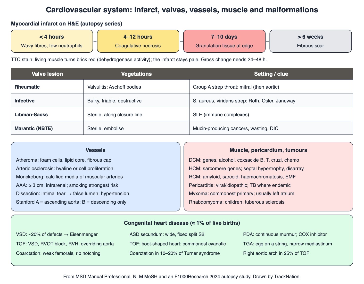
Infarct timeline, valve vegetations, vessels, cardiomyopathy, tumours and congenital heart disease. Drawn by TrackNation.
Arteries and Aneurysms
Arteriosclerosis = arterial wall thickening with lost elasticity. Three forms: atherosclerosis (intimal plaques in large and medium arteries), arteriolosclerosis (small arteries and arterioles thickened by cell proliferation or hyaline deposition) and Mönckeberg medial calcific sclerosis (calcified media of muscular arteries).
Atheroma: monocytes become macrophages, take up lipoprotein through scavenger receptors and turn into foam cells — the hallmark of the early plaque. A mature plaque has a lipid core under a fibrous cap of smooth muscle and collagen. A vulnerable plaque has a thin cap and a big lipid core and is the one that ruptures.
Abdominal aortic aneurysm: diameter ≥ 3 cm, usually infrarenal. Smoking is the strongest risk factor. Repair when > 5.5 cm in men or > 5 cm in women. Mycotic (infected) aneurysm: S. aureus first, then Salmonella.
Aortic dissection: blood surges through an intimal tear and splits intima from media, making a false lumen. Hypertension is a key contributor. Peak age 50–65 in general but 20–40 in Marfan or Ehlers-Danlos. Stanford A involves the ascending aorta; B is confined to the descending aorta beyond the left subclavian.
Primary (essential) hypertension is about 85% of cases; the rest are secondary (for example primary aldosteronism, renal disease).
Ischaemic Heart Disease
Spectrum: unstable angina (ischaemia, troponin below the 99th percentile) → NSTEMI (subendocardial; troponin up, no ST elevation) → STEMI. MI = troponin rise above the 99th percentile plus ischaemic symptoms, ECG change or imaging evidence.
Type 1 MI: plaque rupture, erosion or fissuring with thrombus. Type 2: supply-demand mismatch (for example coronary spasm or a demand surge). Transmural infarcts span epicardium to endocardium.
Infarct age on histology (autopsy series): < 4 h wavy fibres with a few neutrophils; 4–12 h coagulative necrosis; 7–10 days granulation tissue at the edge; > 6 weeks fibrous scar. Gross changes need 24–48 h, so very early infarcts are found with TTC stain: living muscle turns brick red (dehydrogenase activity); the infarct stays pale.
Site clues: right ventricular infarction usually means proximal right coronary artery occlusion. Papillary muscle dysfunction gives a soft apical murmur of mitral regurgitation; a friction rub on days 2–3 after STEMI is common.
Rheumatic Fever and Endocarditis
Rheumatic fever: a non-suppurative complication of group A streptococcal pharyngitis — arthritis, carditis, subcutaneous nodules, erythema marginatum, chorea (modified Jones criteria). Evidence of preceding infection: throat culture/antigen or ASO > 320 U.
Aschoff bodies (granulomatous collections of leukocytes, myocytes and collagen) form in the myocardium. Carditis is a pancarditis, but valvulitis is its most consistent feature. Acute: mitral regurgitation; chronic rheumatic heart disease: mitral stenosis, then aortic disease. Sydenham chorea reflects basal ganglia involvement.
Infective endocarditis: S. aureus attacks even normal valves; viridans streptococci follow dental bacteraemia (even toothbrushing with gingivitis). Mostly left-sided; 5–10% right-sided (tricuspid, pulmonary), much commoner in IV drug users. HACEK organisms are a minority. Signs: Roth spots (retina), Osler nodes (painful, finger tips), Janeway lesions (painless, palms and soles), splinter haemorrhages.
Valve lesion | Vegetations | Setting / clue |
Rheumatic | Valvulitis with Aschoff bodies in the myocardium | Group A streptococcal throat infection; mitral, then aortic valve |
Infective | Infected, friable; destroy valves, cause emboli | S. aureus (even normal valves), viridans streptococci after dental bacteraemia |
Libman-Sacks | Sterile platelet-fibrin, along the leaflet closure line | SLE (circulating immune complexes) |
Marantic (non-bacterial thrombotic) | Sterile, embolise | Mucin-producing cancers (lung, stomach, pancreas), wasting disease, DIC |
Myocardium, Pericardium and Tumours
Dilated cardiomyopathy: genetic, or acquired — coxsackievirus B, Trypanosoma cruzi, alcohol, cardiotoxic chemotherapy. The commonest cardiomyopathy of childhood.
Hypertrophic cardiomyopathy: usually a sarcomere gene mutation. Asymmetric hypertrophy of the basal septum; histology shows myocyte hypertrophy, myocyte disarray and fibrosis. Systolic murmur louder with Valsalva; syncope and sudden death.
Restrictive cardiomyopathy: stiff walls that impede filling with near-normal systolic function — amyloidosis, sarcoidosis, haemochromatosis, Fabry, and endomyocardial fibrosis (children and young adults in the tropics).
Pericarditis: mostly viral or idiopathic in the West, TB where TB is endemic; also uraemia, MI, cancer. Constrictive pericarditis = fibrous scarring fuses both layers. Tamponade = compression by fluid or blood.
Cardiac tumours: metastases are far commoner than primaries. Myxoma is the commonest primary, usually in the left atrium; about 15% of pedunculated ones give a "tumour plop". Rhabdomyoma is the commonest in children — up to 90% have tuberous sclerosis.
Congenital Heart Disease
The commonest congenital anomaly, in almost 1% of live births.
VSD: about 20% of defects; loud harsh holosystolic murmur at the lower left sternal border. A large shunt raises pulmonary resistance until flow reverses — Eisenmenger syndrome.
ASD (secundum, at the fossa ovalis): wide, fixed splitting of S2; paradoxical emboli later. PDA: continuous murmur and bounding pulses; a COX inhibitor (ibuprofen or indomethacin) may close it in preterm babies.
Tetralogy of Fallot — the commonest cyanotic lesion (twice TGA): large VSD, RV outflow obstruction, RV hypertrophy, overriding aorta. Boot-shaped heart; right aortic arch in 25%. TGA: egg-on-a-string shadow with a narrow mediastinum.
Coarctation: in 10–20% of Turner syndrome; arm hypertension with weak, delayed femoral pulses; collateral intercostal arteries notch ribs 3–8 (rarely before age 5).
Traps the Examiner Sets
Osler = Ouch (painful); Janeway = painless.
Libman-Sacks (SLE) and marantic vegetations are sterile; infective ones are not.
Acute rheumatic carditis → mitral regurgitation; chronic → mitral stenosis.
TTC: pale = dead; brick red = alive.
Commonest primary cardiac tumour: myxoma (adult, left atrium); in children: rhabdomyoma (tuberous sclerosis).
Memory Hooks & Shortcuts
Shortcuts to lock in this article's numbers and lists. Each one only re-packages facts proven above.
🧠 Osler = Ouch; Janeway = painless (J for 'just a spot') — IE skin signs
🧠 Wavy at 4, Necrosis by 12, Granulation at 10 days, Scar at 6 weeks — infarct dating
🧠 TTC: Tissue That's Caput stays pale — TTC staining
🧠 Libman-Sacks = Lupus; Marantic = Malignancy (mucin) — sterile vegetations
🧠 Myxoma Mostly in the left atriuM — myxoma
🧠 Rhabdo for babies with tubers (tuberous sclerosis) — rhabdomyoma
🧠 Turner turns the aorta tight (coarctation) — coarctation
Rapid-Revision Cards
Arteriolosclerosis
Small arteries/arterioles: hyaline or cell proliferation.
Mönckeberg
Calcified media of muscular arteries.
Early plaque hallmark
Foam cells (lipid-laden macrophages).
Vulnerable plaque
Thin fibrous cap, big lipid core.
AAA
≥ 3 cm, infrarenal; smoking; repair > 5.5 cm (men).
Mycotic aneurysm
S. aureus, then Salmonella.
Stanford A vs B
A = ascending aorta; B = descending only.
Infarct < 4 h
Wavy fibres, few neutrophils.
Infarct 4–12 h
Coagulative necrosis.
Infarct 7–10 d
Granulation tissue at the edge.
Infarct > 6 wk
Fibrous scar.
TTC stain
Brick red = alive; pale = infarct.
Type 1 vs 2 MI
1 = plaque rupture; 2 = supply-demand.
Aschoff body
Rheumatic carditis; myocardium.
Rheumatic valve
Acute MR; chronic mitral stenosis.
ASO in ARF
> 320 U.
Osler vs Janeway
Osler painful finger tips; Janeway painless palms/soles.
IV drug user IE
Right-sided (tricuspid).
Libman-Sacks
SLE; sterile; closure line.
Marantic
Mucin-producing cancer; sterile; embolise.
HCM
Sarcomere genes; septum; disarray; Valsalva ↑ murmur.
RCM causes
Amyloid, sarcoid, haemochromatosis, EMF.
Myxoma
Commonest primary; left atrium; tumour plop.
Rhabdomyoma
Children; tuberous sclerosis.
TOF
VSD, RVOT block, RVH, overriding aorta; boot heart.
ASD
Wide fixed split S2.
Coarctation
Turner; weak femorals; rib notching.
Sources
Facts in this article were checked against these documents.
MSD Manual Professional: Atherosclerosis; Abdominal Aortic Aneurysms; Aortic Dissection; Hypertension; Heart Failure — Merck & Co. (MSD Manuals), reviewed 2024–26 (link)
MSD Manual Professional: Acute Coronary Syndromes; Acute Myocardial Infarction; Infective and Noninfective Endocarditis; Rheumatic Fever; Dilated, Hypertrophic and Restrictive Cardiomyopathy; Pericarditis; Cardiac Tumors — Merck & Co. (MSD Manuals), reviewed 2025–26 (link)
MSD Manual Professional: Congenital Cardiovascular Anomalies (overview, VSD, ASD, PDA, Tetralogy of Fallot, TGA, Coarctation) — Merck & Co. (MSD Manuals), reviewed 2023–26 (link)
Standard Treatment Workflow: Acute Rheumatic Fever — ICMR, 2022
Medical Subject Headings (scope notes) — US National Library of Medicine, 2026 (link)
Varun KB et al. Comparison of histopathological and triphenyl tetrazolium chloride test in diagnosing myocardial infarction: an autopsy study. F1000Research (CC BY 4.0) — F1000Research, 2024 (link)
Competency Based Undergraduate Curriculum, Pathology (PA 26) — National Medical Commission, 2024
Test Yourself: Practice Questions
Thirty-four questions in NEET PG style, most of them clinical vignettes.
Practice Questions (NEET PG pattern)
Original questions written in the style of the exam: +4 for a correct answer, −1 for a wrong one. Every answer was checked against the sources listed above. NEET PG papers are not published by NBEMS, so these are not past-paper questions.
Q1. A kidney biopsy shows small arteries and arterioles whose walls are thickened by hyaline deposition. This lesion is
(a) fibromuscular dysplasia
(b) arteriolosclerosis
(c) Mönckeberg medial calcific sclerosis
(d) atherosclerosis
Show answer
Answer: (b) — Arteriolosclerosis thickens small vessels by hyaline deposition or cell proliferation.
Q2. Sections of a muscular artery show calcification confined to the tunica media, with the intima spared. The condition is
(a) Buerger disease
(b) atherosclerosis
(c) arteriolosclerosis
(d) Mönckeberg medial calcific sclerosis
Show answer
Answer: (d) — Calcification of the tunica media of muscular arteries.
Q3. The cell that is the hallmark of the early atherosclerotic plaque is the
(a) plasma cell
(b) Langhans giant cell
(c) eosinophil
(d) foam cell (lipid-laden macrophage)
Show answer
Answer: (d) — Macrophages ingest lipoprotein through scavenger receptors.
Q4. Which plaque is most likely to rupture and cause an acute coronary event?
(a) a fatty dot without a core
(b) heavily calcified small plaque
(c) thick fibrous cap with a small lipid core
(d) thin fibrous cap with a large lipid core
Show answer
Answer: (d) — Matrix breakdown thins and weakens the cap.
Q5. A 72-year-old smoker has a pulsatile abdominal mass. Ultrasound shows an infrarenal aortic diameter of 3.8 cm. The single strongest risk factor for this lesion is
(a) syphilis
(b) diabetes
(c) female sex
(d) smoking
Show answer
Answer: (d) — AAA = aortic diameter ≥ 3 cm.
Q6. The organism most often responsible for a mycotic (infected) aortic aneurysm is
(a) Candida
(b) Staphylococcus aureus
(c) Streptococcus pyogenes
(d) Treponema pallidum
Show answer
Answer: (b) — Salmonella is second.
Q7. A 30-year-old with Marfan syndrome has abrupt tearing chest pain. Imaging shows an intimal flap and double lumen involving the ascending aorta. This is
(a) Stanford type A aortic dissection
(b) abdominal aortic aneurysm
(c) Stanford type B dissection
(d) pulmonary embolism
Show answer
Answer: (a) — Type A involves the ascending aorta; connective tissue disorders lower the age of onset.
Q8. The basic lesion of aortic dissection is
(a) a saccular outpouching of all three layers
(b) an intimal tear with blood splitting intima from media (false lumen)
(c) a full-thickness wall rupture
(d) medial calcification
Show answer
Answer: (b) — Hypertension is an important contributor.
Q9. A man with severe chest pain dies within 3 hours. The most likely microscopic finding in the infarcted myocardium is
(a) wavy fibres with a few neutrophils
(b) dense fibrous scar
(c) granulation tissue
(d) macrophages full of haemosiderin only
Show answer
Answer: (a) — Coagulative necrosis is obvious from about 4–12 hours.
Q10. At autopsy, the margin of a myocardial infarct shows well-formed granulation tissue. The infarct is most likely about
(a) 2 hours old
(b) 7–10 days old
(c) 12 hours old
(d) 6 months old
Show answer
Answer: (b) — A scar is seen after 6 weeks.
Q11. A heart slice is immersed in triphenyl tetrazolium chloride (TTC). A pale unstained area against brick-red muscle means
(a) normal myocardium
(b) fatty infiltration
(c) infarcted myocardium that has lost dehydrogenase activity
(d) fixation artefact
Show answer
Answer: (c) — Useful because gross infarct changes need 24–48 hours.
Q12. An MI caused by coronary plaque rupture with thrombosis is classified as
(a) type 3
(b) type 2
(c) type 5
(d) type 1
Show answer
Answer: (d) — Type 2 is supply-demand mismatch.
Q13. A patient has rest angina for 30 minutes; ECG has no ST elevation and troponin stays below the 99th percentile. The diagnosis is
(a) unstable angina
(b) STEMI
(c) NSTEMI
(d) type 2 MI
Show answer
Answer: (a) — No necrosis means no troponin rise.
Q14. An infarct of the right ventricle most often results from occlusion of the
(a) left anterior descending artery
(b) left main coronary artery
(c) diagonal branch
(d) proximal right coronary artery
Show answer
Answer: (d) — Raised RV filling pressure, often with tricuspid regurgitation.
Q15. Two to three weeks after a sore throat, a child develops migratory polyarthritis and an apical pansystolic murmur. Which antibody titre supports preceding streptococcal infection?
(a) anti-dsDNA
(b) anti-CCP
(c) ANCA
(d) ASO above 320 units
Show answer
Answer: (d) — Or a positive throat culture or rapid antigen test.
Q16. Heart tissue from a child who died of acute rheumatic carditis shows granulomatous collections of leukocytes, myocytes and collagen. These are
(a) Aschoff bodies
(b) Anitschkow cells alone
(c) Mallory bodies
(d) Langhans giant cells
Show answer
Answer: (a) — They develop in the myocardium and other parts of the heart.
Q17. The commonest valve lesion of chronic rheumatic heart disease is
(a) mitral stenosis
(b) tricuspid stenosis alone
(c) pulmonary stenosis
(d) aortic coarctation
Show answer
Answer: (a) — Acute carditis more often gives mitral regurgitation.
Q18. The most consistent feature of rheumatic carditis, without which the diagnosis should be reconsidered, is
(a) valvulitis
(b) myocardial abscess
(c) heart block
(d) pericardial effusion
Show answer
Answer: (a) — Although it is a pancarditis.
Q19. A patient with fever and a murmur has painful red nodules on the finger pads. These are
(a) Osler nodes
(b) splinter haemorrhages
(c) Janeway lesions
(d) Roth spots
Show answer
Answer: (a) — Janeway lesions are painless and on palms and soles.
Q20. A person who injects drugs has septic pulmonary emboli. The valve most likely involved by endocarditis is the
(a) tricuspid valve
(b) mitral valve
(c) a prosthetic mitral valve
(d) aortic valve
Show answer
Answer: (a) — Right-sided disease is much commoner in IV drug users.
Q21. An organism able to cause endocarditis on a previously normal valve is
(a) Staphylococcus aureus
(b) Lactobacillus
(c) Staphylococcus epidermidis on a native valve
(d) Corynebacterium diphtheriae
Show answer
Answer: (a) — Massive bacteraemia or virulent organisms.
Q22. A woman with SLE has small sterile vegetations along the closure line of the mitral leaflets. These are
(a) Libman-Sacks lesions
(b) marantic vegetations
(c) Lambl excrescences
(d) infective vegetations
Show answer
Answer: (a) — Immune complexes cause platelet-fibrin vegetations.
Q23. A man with metastatic pancreatic adenocarcinoma has a systemic arterial embolus; echo shows valve vegetations and blood cultures are negative. The lesion is
(a) Libman-Sacks endocarditis
(b) rheumatic valvulitis
(c) carcinoid heart disease
(d) marantic (non-bacterial thrombotic) endocarditis
Show answer
Answer: (d) — Mucin-producing carcinomas of lung, stomach or pancreas.
Q24. A young athlete collapses and dies. Autopsy shows asymmetric thickening of the basal interventricular septum and microscopic myocyte disarray. The usual cause is
(a) a sarcomere protein gene mutation
(b) amyloid deposition
(c) alcohol
(d) coxsackievirus myocarditis
Show answer
Answer: (a) — Hypertrophic cardiomyopathy; murmur louder with Valsalva.
Q25. Which of these is a recognised infectious cause of dilated cardiomyopathy?
(a) Mycobacterium leprae
(b) Treponema pallidum
(c) Trypanosoma cruzi
(d) Entamoeba histolytica
Show answer
Answer: (c) — Also coxsackievirus B; alcohol and chemotherapy are toxic causes.
Q26. Restrictive cardiomyopathy with stiff ventricles and near-normal systolic function is classically caused by
(a) sarcomere gene mutation
(b) coxsackievirus B
(c) chronic alcohol use
(d) amyloidosis
Show answer
Answer: (d) — Also sarcoidosis, haemochromatosis, Fabry disease.
Q27. In a country where tuberculosis is endemic, the commonest cause of pericarditis is
(a) tuberculosis
(b) uraemia
(c) radiation
(d) viral infection
Show answer
Answer: (a) — In the West it is mostly viral or idiopathic.
Q28. A woman has a murmur like mitral stenosis whose loudness varies with body position; echo shows a pedunculated left atrial mass. The likely tumour is
(a) rhabdomyoma
(b) fibroma
(c) myxoma
(d) angiosarcoma
Show answer
Answer: (c) — The commonest primary cardiac tumour; most arise in the left atrium.
Q29. An infant with multiple cardiac tumours in the ventricular walls is most likely to have which underlying syndrome?
(a) tuberous sclerosis
(b) Down syndrome
(c) Marfan syndrome
(d) Turner syndrome
Show answer
Answer: (a) — Rhabdomyoma, the commonest cardiac tumour of children.
Q30. A cyanotic infant's chest X-ray shows a boot-shaped heart with reduced pulmonary markings. The four components of the lesion are
(a) ASD, PDA, LVH, coarctation
(b) VSD, ASD, PDA, TGA
(c) mitral stenosis, AR, TR, PS
(d) VSD, RV outflow obstruction, RV hypertrophy, overriding aorta
Show answer
Answer: (d) — Tetralogy of Fallot, the commonest cyanotic congenital heart disease.
Q31. A child with an uncorrected large VSD later becomes cyanosed as pulmonary resistance rises and the shunt reverses. This is
(a) Eisenmenger syndrome
(b) Ebstein anomaly
(c) tetralogy of Fallot
(d) transposition
Show answer
Answer: (a) — Right-to-left shunting.
Q32. Wide and fixed splitting of the second heart sound is typical of
(a) patent ductus arteriosus
(b) ventricular septal defect
(c) coarctation
(d) atrial septal defect
Show answer
Answer: (d) — Secundum ASD lies at the fossa ovalis.
Q33. A preterm infant has bounding pulses and a continuous murmur at the upper left sternal border. Medical closure is attempted with
(a) a cyclo-oxygenase inhibitor such as ibuprofen or indomethacin
(b) a beta-blocker
(c) digoxin
(d) prostaglandin E1
Show answer
Answer: (a) — Patent ductus arteriosus; after birth, falling prostaglandin normally closes the duct.
Q34. A girl with Turner syndrome has hypertension in the arms with weak, delayed femoral pulses. The likely cardiovascular lesion is
(a) coarctation of the aorta
(b) Ebstein anomaly
(c) tetralogy of Fallot
(d) patent ductus arteriosus
Show answer
Answer: (a) — Occurs in 10–20% of Turner syndrome; rib notching may appear later.
Active Recall
From memory: distinguish the three forms of arteriosclerosis; describe the atheroma and vulnerable plaque; define AAA and dissection; date an infarct from histology and explain TTC staining; describe rheumatic fever, Aschoff bodies and its valve lesions; compare infective, Libman-Sacks and marantic vegetations; summarise DCM, HCM and RCM; name the commonest cardiac tumours; and give the clues for VSD, ASD, PDA, TOF and coarctation.