Kidney I for NEET PG Pathology: AKI, CKD, Nephritic and Nephrotic Syndrome, Glomerulonephritis, IgA, RPGN, Lupus and Diabetic Nephropathy
Start Here
🎯 Exam priority: High. Expect: nephritic vs nephrotic, the biopsy hallmark of each glomerular disease (foot processes, spikes, humps, linear IgG, crescents, Kimmelstiel-Wilson), IgA vs post-infectious timing, lupus classes, AKI criteria and prerenal vs tubular indices, and the casts of CKD.
CBME mapping: PA 27.2–27.7. Tubules, infection, cysts, stones and tumours are in article 22; amyloid kidney in article 6; urine examination in article 16.
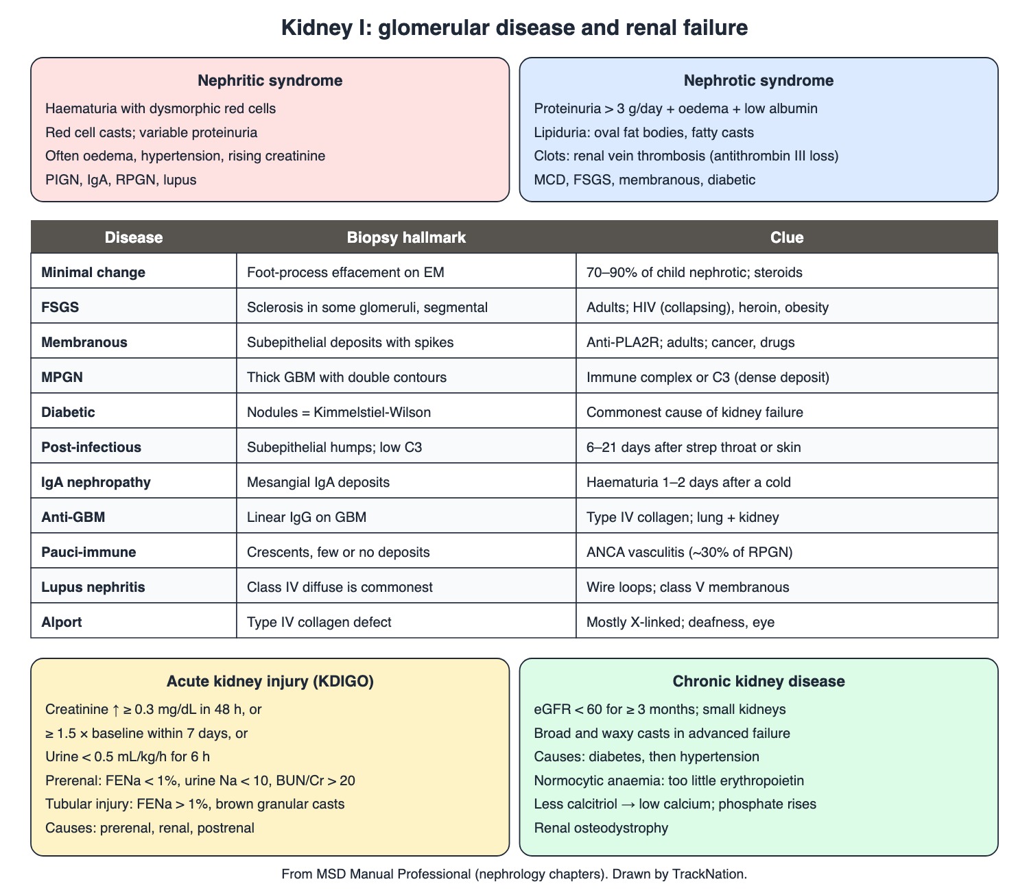
Nephritic vs nephrotic syndrome, glomerular diseases, AKI and CKD. Drawn by TrackNation.
Acute and Chronic Kidney Injury
AKI: a fall in kidney function over days to weeks with azotaemia. KDIGO definition — creatinine up ≥ 0.3 mg/dL within 48 hours, or ≥ 1.5 times baseline within 7 days, or urine < 0.5 mL/kg/h for 6 hours. Causes are prerenal (poor perfusion), renal (intrinsic) or postrenal (obstruction). Prerenal AKI and acute tubular injury are the commonest in hospital.
Tubular injury gives tubular cells and many brown granular casts; urinary eosinophils hint at allergic interstitial nephritis.
CKD: eGFR < 60 mL/min/1.73 m² for ≥ 3 months is the most reliable evidence; small kidneys on ultrasound favour CKD. Commonest causes: diabetic nephropathy, then hypertensive nephrosclerosis. Broad and waxy casts mark advanced failure.
CKD complications: normocytic normochromic anaemia (too little erythropoietin); less calcitriol → low calcium, phosphate retention, secondary hyperparathyroidism and renal osteodystrophy.
Index | Prerenal | Tubular injury |
Urine sodium (mmol/L) | < 10 | > 40 |
Fractional excretion of sodium | < 1% | > 1% |
BUN/creatinine ratio | > 20 | < 10 |
Urine/plasma osmolality | > 1.5 | 1–1.5 |
Sediment | Bland | Tubular cells, many brown granular casts |
Nephrotic Syndrome
Definition: protein > 3 g/day from a glomerular disorder, with oedema and hypoalbuminaemia. Lipiduria (oval fat bodies, fatty casts). Loss of antithrombin III → clots, especially renal vein thrombosis.
Minimal change disease: 70–90% of nephrotic syndrome in children over 1 year; diffuse effacement of foot processes on electron microscopy; steroid responsive. Rare secondary causes: NSAIDs, Hodgkin lymphoma.
FSGS: sclerosis begins in some (focal) glomeruli and in part of each tuft (segmental); commonest primary nephrotic syndrome in US adults. Secondary: heroin, HIV, obesity, sickle cell disease. HIV-associated nephropathy shows the collapsing variant.
Membranous nephropathy: an adult disease; target antigen PLA2R on podocytes. Subepithelial deposits with spikes of basement membrane between them. Secondary to drugs, infections, autoimmunity, cancer.
MPGN: thickened GBM with double contours; immune-complex or complement-mediated (C3 GN, dense deposit disease).
Diabetic nephropathy: commonest cause of nephrotic syndrome in adults and of kidney failure worldwide. Early hyperfiltration; thick GBM; nodular glomerulosclerosis = Kimmelstiel-Wilson lesions; hyalinosis of both afferent and efferent arterioles.
Nephritic Syndrome and RPGN
Definition: haematuria with dysmorphic red cells and often red cell casts, variable proteinuria.
Post-infectious GN: commonest acute GN in children 3–15 years; nephritogenic group A streptococci (pharyngitis or impetigo strains) after a latency of 6–21 days. Cola-coloured urine, oedema, hypertension. Subepithelial humps on EM; low C3 that normalises within 6–8 weeks in 80%.
IgA nephropathy: commonest GN worldwide. Poorly glycosylated IgA1 binds mesangial cells. Gross haematuria 1–2 days after a mucosal infection — a shorter latency than post-infectious GN.
RPGN: nephritic syndrome with glomerular crescents and kidney failure in weeks to months. By immunofluorescence: anti-GBM (~20%, linear IgG; Goodpasture when the lung bleeds too — antibody against type IV collagen), immune complex (~50%; post-infectious, IgA, lupus and others) and pauci-immune (~30%, ANCA vasculitis such as granulomatosis with polyangiitis).
Lupus nephritis: class I minimal, II mesangial, III focal (< 50% glomeruli), IV diffuse (> 50%, commonest), V membranous (nephrotic), VI sclerosing. Wire loops are an activity lesion.
Hereditary: Alport syndrome (type IV collagen gene, mostly X-linked, with sensorineural deafness and eye changes); thin basement membrane disease (GBM 150–225 nm instead of 300–400 nm; isolated microscopic haematuria).
Traps the Examiner Sets
Haematuria 1–2 days after a cold = IgA; 1–3 weeks after a sore throat or skin infection = post-infectious GN.
Linear IgG = anti-GBM; immune-complex deposits = immune-complex GN; few or none = pauci-immune (ANCA).
Spikes = membranous; humps = post-infectious; foot processes only = minimal change.
FENa < 1% = prerenal; > 1% = tubular injury.
Waxy and broad casts = advanced CKD; red cell casts = glomerulonephritis.
Memory Hooks & Shortcuts
Shortcuts to lock in this article's numbers and lists. Each one only re-packages facts proven above.
🧠 IgA comes In A day (1–2 days); strep takes three weeks — IgA vs PIGN latency
🧠 Spikes for Membranous, Humps for Post-strep, Foot processes for Minimal change — EM hallmarks
🧠 Linear = Lung + kidney (anti-GBM) — Goodpasture
🧠 Kimmelstiel-Wilson = Kandy (diabetes) nodules — diabetic nephropathy
🧠 Waxy = Way past it (advanced CKD) — casts
🧠 Prerenal Preserves sodium (FENa < 1%) — AKI indices
Rapid-Revision Cards
AKI (KDIGO)
Cr ↑ ≥ 0.3 mg/dL in 48 h, or ≥ 1.5× in 7 d, or urine < 0.5 mL/kg/h × 6 h.
Prerenal indices
Urine Na < 10, FENa < 1%, BUN/Cr > 20.
Tubular injury
FENa > 1%; brown granular casts.
CKD
eGFR < 60 for ≥ 3 months; small kidneys.
CKD casts
Broad and waxy.
CKD anaemia
Normocytic; low erythropoietin.
CKD calcium
Low calcitriol → hypocalcaemia.
Nephrotic
> 3 g/day, oedema, low albumin, lipiduria.
Nephrotic clot
Renal vein thrombosis; AT-III loss.
Minimal change
Children; foot-process effacement; steroids; Hodgkin.
FSGS
Adults (US); heroin, HIV, obesity, sickle.
HIVAN
Collapsing FSGS.
Membranous
Adults; PLA2R; subepithelial spikes.
MPGN
Double-contour GBM.
Diabetic kidney
Kimmelstiel-Wilson nodules; afferent + efferent hyalinosis.
Nephritic
Haematuria, dysmorphic RBCs, RBC casts.
PIGN
6–21 days after strep; humps; low C3.
IgA nephropathy
Commonest GN; haematuria 1–2 days after URTI.
RPGN
Crescents; anti-GBM 20%, IC 50%, pauci 30%.
Anti-GBM
Linear IgG; type IV collagen; lung + kidney.
Lupus nephritis
Class IV diffuse commonest; V membranous.
Alport
Type IV collagen; X-linked; deafness.
Thin GBM
150–225 nm; benign haematuria.
Sources
Facts in this article were checked against these documents.
MSD Manual Professional: Acute Kidney Injury; Chronic Kidney Disease — Merck & Co. (MSD Manuals), reviewed 2026 (link)
MSD Manual Professional: Glomerular Disorders (nephritic and nephrotic overviews, PIGN, IgA nephropathy, RPGN, minimal change disease, FSGS, membranous nephropathy, MPGN, diabetic nephropathy, lupus nephritis, Alport syndrome, thin basement membrane disease, HIV-associated nephropathy) — Merck & Co. (MSD Manuals), reviewed 2025–26 (link)
MSD Manual Professional: Anti-Glomerular Basement Membrane Disease — Merck & Co. (MSD Manuals), reviewed 2025 (link)
Competency Based Undergraduate Curriculum, Pathology (PA 27) — National Medical Commission, 2024
Test Yourself: Practice Questions
Thirty-two questions in NEET PG style, most of them clinical vignettes.
Practice Questions (NEET PG pattern)
Original questions written in the style of the exam: +4 for a correct answer, −1 for a wrong one. Every answer was checked against the sources listed above. NEET PG papers are not published by NBEMS, so these are not past-paper questions.
Q1. A hospital patient's creatinine rises from 1.0 to 1.4 mg/dL within 48 hours. By KDIGO criteria this is
(a) normal variation
(b) chronic kidney disease
(c) nephrotic syndrome
(d) acute kidney injury
Show answer
Answer: (d) — A rise of ≥ 0.3 mg/dL in 48 hours meets the definition.
Q2. An oliguric patient after severe diarrhoea has urine sodium 8 mmol/L, FENa 0.5% and BUN/creatinine ratio 25. The AKI is most likely
(a) acute tubular injury
(b) postrenal obstruction
(c) prerenal
(d) acute interstitial nephritis
Show answer
Answer: (c) — Tubular injury: urine Na > 40, FENa > 1%, ratio < 10.
Q3. Urine sediment shows tubular epithelial cells and many brown granular casts in an oliguric patient after prolonged hypotension. This points to
(a) IgA nephropathy
(b) prerenal azotaemia
(c) minimal change disease
(d) acute tubular injury
Show answer
Answer: (d) — Prerenal sediment is usually bland.
Q4. The most reliable evidence that kidney disease is chronic rather than acute is
(a) eGFR below 60 for at least 3 months
(b) oliguria
(c) a single high creatinine
(d) red cell casts
Show answer
Answer: (a) — Small kidneys on ultrasound also favour CKD.
Q5. Urine from a patient with advanced renal failure shows broad, highly refractile casts. They are
(a) white cell casts
(b) waxy casts
(c) red cell casts
(d) hyaline casts only
Show answer
Answer: (b) — Broad or waxy casts are prominent in advanced failure of any cause.
Q6. A patient with stage 4 CKD has haematocrit 25%, normal MCV and normal MCHC. The main cause is
(a) iron deficiency always
(b) haemolysis
(c) deficient erythropoietin production
(d) B12 deficiency
Show answer
Answer: (c) — Anaemia of CKD is normochromic normocytic.
Q7. Hypocalcaemia in CKD is mainly due to
(a) excess vitamin D
(b) reduced renal production of calcitriol
(c) low phosphate
(d) excess PTH
Show answer
Answer: (b) — Phosphate retention adds to it.
Q8. The two commonest causes of CKD, in order, are
(a) lupus, then IgA
(b) polycystic kidney, then stones
(c) TB, then amyloid
(d) diabetic nephropathy, then hypertensive nephrosclerosis
Show answer
Answer: (d) — Then glomerulopathies.
Q9. A 4-year-old has oedema, urine protein over 3 g/day, a non-nephritic sediment and normal kidney function. Electron microscopy shows diffuse effacement of podocyte foot processes. The diagnosis is
(a) post-infectious GN
(b) membranous nephropathy
(c) Alport syndrome
(d) minimal change disease
Show answer
Answer: (d) — 70–90% of nephrotic syndrome in children over 1 year; steroid responsive.
Q10. A patient with Hodgkin lymphoma develops nephrotic syndrome. The glomerular lesion classically associated is
(a) anti-GBM disease
(b) membranous nephropathy
(c) IgA nephropathy
(d) minimal change disease
Show answer
Answer: (d) — NSAIDs are the other secondary cause.
Q11. Nephrotic syndrome is defined by proteinuria above
(a) 1 g/day
(b) 30 mg/g creatinine
(c) 150 mg/day
(d) 3 g/day with oedema and hypoalbuminaemia
Show answer
Answer: (d) — From a glomerular disorder.
Q12. The thrombotic complication especially typical of nephrotic syndrome is
(a) renal vein thrombosis
(b) papillary necrosis
(c) pyelonephritis
(d) renal stone
Show answer
Answer: (a) — Antithrombin III is lost in the urine.
Q13. Urine microscopy in nephrotic syndrome shows tubular cells packed with lipid. These are
(a) clue cells
(b) red cell casts
(c) waxy casts
(d) oval fat bodies
Show answer
Answer: (d) — Lipiduria also gives fatty casts.
Q14. Biopsy from an adult with nephrotic syndrome shows subepithelial deposits with spikes of basement membrane between them; serum anti-PLA2R antibody is positive. The diagnosis is
(a) FSGS
(b) membranous nephropathy
(c) MPGN
(d) minimal change disease
Show answer
Answer: (b) — PLA2R on podocytes is the major target antigen.
Q15. A patient with HIV infection and heavy proteinuria has a biopsy showing collapsing glomerulopathy. This is a variant of
(a) IgA nephropathy
(b) focal segmental glomerulosclerosis
(c) membranous nephropathy
(d) MPGN
Show answer
Answer: (b) — HIV-associated nephropathy.
Q16. Secondary FSGS is associated with all of the following EXCEPT
(a) IgA vasculitis as the main cause
(b) obesity
(c) sickle cell disease
(d) heroin use
Show answer
Answer: (a) — Also HIV and nephron loss.
Q17. A silver stain of a glomerulus shows a thickened basement membrane with a double contour ('tram track'). The pattern is
(a) diabetic nephropathy
(b) thin basement membrane disease
(c) minimal change disease
(d) membranoproliferative glomerulonephritis
Show answer
Answer: (d) — Immune-complex or complement (C3) mediated.
Q18. Kidney biopsy from a long-standing diabetic shows rounded nodules of mesangial matrix and hyalinosis of both afferent and efferent arterioles. The nodules are
(a) crescents
(b) wire loops
(c) humps
(d) Kimmelstiel-Wilson lesions
Show answer
Answer: (d) — Nodular intercapillary glomerulosclerosis.
Q19. The commonest cause of kidney failure worldwide is
(a) polycystic kidney disease
(b) IgA nephropathy
(c) diabetic nephropathy
(d) lupus nephritis
Show answer
Answer: (c) — Also the commonest cause of nephrotic syndrome in adults.
Q20. Two weeks after a streptococcal sore throat, a 7-year-old passes cola-coloured urine and has oedema and hypertension. Which finding is expected?
(a) normal complement with IgA deposits
(b) linear IgG on immunofluorescence
(c) positive anti-PLA2R
(d) low serum C3
Show answer
Answer: (d) — C3 returns to normal within 6–8 weeks in 80%.
Q21. Electron microscopy in post-streptococcal glomerulonephritis typically shows
(a) spikes
(b) foot-process effacement only
(c) linear deposits
(d) hump-shaped subepithelial immune complex deposits
Show answer
Answer: (d) — With endocapillary proliferation.
Q22. A man has recurrent gross haematuria; the latest episode began one day after an upper respiratory infection. The likely diagnosis is
(a) post-streptococcal GN
(b) minimal change disease
(c) membranous nephropathy
(d) IgA nephropathy
Show answer
Answer: (d) — Post-infectious GN has a latency of 1–3 weeks.
Q23. The commonest form of glomerulonephritis worldwide is
(a) post-streptococcal GN
(b) IgA nephropathy
(c) anti-GBM disease
(d) lupus nephritis
Show answer
Answer: (b) — Commoner in Asia.
Q24. Rapidly progressive glomerulonephritis is defined histologically by
(a) foot-process effacement
(b) Kimmelstiel-Wilson nodules
(c) thin basement membrane
(d) glomerular crescents
Show answer
Answer: (d) — Kidney failure follows within weeks to months.
Q25. A smoker has haemoptysis and rapidly rising creatinine. Immunofluorescence of the kidney shows linear IgG along the GBM. The antibody targets
(a) neutrophil proteinase 3
(b) PLA2R
(c) double-stranded DNA
(d) type IV collagen of basement membranes
Show answer
Answer: (d) — Anti-GBM (Goodpasture) disease.
Q26. RPGN with crescents but few or no immune deposits on immunofluorescence is usually due to
(a) post-streptococcal GN
(b) ANCA-associated vasculitis
(c) anti-GBM antibody
(d) lupus
Show answer
Answer: (b) — Pauci-immune, about 30% of RPGN.
Q27. The commonest class of lupus nephritis, with proliferation in more than half the glomeruli, is
(a) class VI (sclerosing)
(b) class I (minimal mesangial)
(c) class IV (diffuse)
(d) class II (mesangial)
Show answer
Answer: (c) — Class V is membranous and usually nephrotic.
Q28. A boy has haematuria, progressive kidney disease and high-frequency sensorineural hearing loss; his maternal uncle had kidney failure. The defective protein is
(a) fibrillin
(b) nephrin only
(c) type IV collagen
(d) polycystin
Show answer
Answer: (c) — Alport syndrome, most often X-linked.
Q29. A healthy adult has isolated microscopic haematuria and normal kidney function; electron microscopy shows a GBM about 200 nm wide. The diagnosis is
(a) Alport syndrome
(b) IgA nephropathy
(c) minimal change disease
(d) thin basement membrane disease
Show answer
Answer: (d) — Normal GBM is 300–400 nm.
Q30. Nephritic syndrome is characterised by
(a) proteinuria > 3 g/day with lipiduria
(b) glycosuria
(c) haematuria with dysmorphic red cells and red cell casts
(d) pyuria with white cell casts
Show answer
Answer: (c) — Proteinuria is variable.
Q31. Membranous nephropathy is best described as
(a) a crescentic GN
(b) mainly an adult disease, often secondary to drugs, infections, autoimmunity or cancer
(c) the commonest nephrotic syndrome of young children
(d) a hereditary collagen disease
Show answer
Answer: (b) — Most cases are primary (PLA2R).
Q32. The commonest cause of primary nephrotic syndrome in adults (US data) is
(a) post-infectious GN
(b) focal segmental glomerulosclerosis
(c) IgA nephropathy
(d) minimal change disease
Show answer
Answer: (b) — Especially common in Black men.
Active Recall
From memory: define AKI by KDIGO and separate prerenal from tubular injury; define CKD and list its casts, anaemia and mineral changes; contrast nephritic and nephrotic syndrome; give the biopsy hallmark of minimal change, FSGS, membranous, MPGN and diabetic nephropathy; contrast IgA and post-infectious GN; classify RPGN by immunofluorescence; and name the commonest lupus class and the Alport defect.