Fluids, Electrolytes and Acid–Base for NEET PG Medicine: Hyponatraemia (ICMR), Potassium, Calcium, Anion Gap
Start Here
🎯 Exam priority: High. Expect: hyponatraemia by volume status, 3% saline and the 8–10 mmol/L limit, SIADH criteria, potassium ECG changes, pseudohyperkalaemia, Chvostek and Trousseau, hypercalcaemia treatment, magnesium with low potassium, and high vs normal anion gap acidosis.
CBME mapping: GM 23.1–23.12. Hyperkalaemia emergency treatment is also in article 15; DKA acid–base in article 17; renal physiology in Physiology.
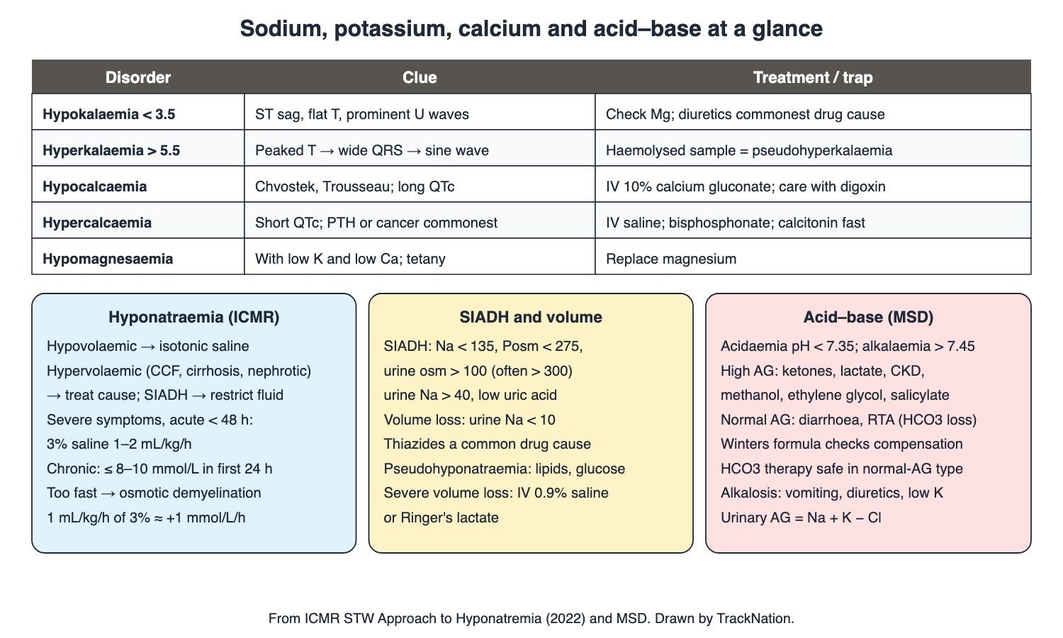
Electrolyte disorders, ICMR hyponatraemia approach, SIADH and acid–base. Drawn by TrackNation.
Volume and Sodium
Volume depletion: reduced skin turgor, dry mucosa, tachycardia, orthostatic hypotension; urine sodium < 10. Severe: IV 0.9% saline or Ringer's lactate.
Hyponatraemia causes: diuretics (especially thiazides), diarrhoea, heart failure, liver and kidney disease, SIADH. Severe if < 125 mmol/L. Pseudohyponatraemia is caused by glucose or lipids, not a water imbalance.
SIADH: Na < 135, plasma osmolality < 275, urine osmolality > 100 (often > 300), urine Na > 40, low uric acid. Euvolaemic. Restrict fluid; oral tolvaptan 15 mg on day 1 (ICMR) if Na stays low.
Hypernatraemia: replace the intravascular volume and the free-water deficit.
Hyponatraemia (ICMR STW) | Action |
Hypovolaemic | IV isotonic normal saline |
Hypervolaemic (heart failure, cirrhosis, nephrotic) | Treat the underlying disease |
Severe symptoms (vomiting, seizures, coma) and acute < 48 h | Emergency 3% saline 1–2 mL/kg/h to a safe level (a 4–6 mmol/L rise relieves symptoms) |
Chronic or unknown (> 48 h) | 3% saline 0.5–1 mL/kg/h only for symptoms; restrict fluid to 500–1000 mL/day |
Correction limit | Not more than 8–10 mmol/L in the first 24 h — faster risks osmotic demyelination (central pontine myelinolysis) |
Arithmetic | 3% saline = 513 mmol Na/L; 1 mL/kg/h raises Na by ~1 mmol/L/h; 0.9% saline = 154 mmol/L |
Potassium, Calcium, Magnesium
Hypokalaemia (< 3.5): ST sagging, T-wave depression, prominent U waves. Diuretics are the commonest drug cause; low magnesium increases renal K loss — correct it. Replace with oral KCl (IV if severe).
Hyperkalaemia (> 5.5): peaked T waves → (> 6.5) wide QRS → sine wave → VF/asystole. A haemolysed sample gives pseudohyperkalaemia.
Hypocalcaemia: hypoparathyroidism (low Ca, high phosphate), vitamin D deficiency, renal disease. Chvostek (facial twitch on tapping in front of the ear) and Trousseau (carpal spasm with cuff 20 mm Hg above systolic for 3 min); prolonged QTc. Severe: IV 10% calcium gluconate — hazardous with digoxin.
Hypercalcaemia: commonest causes hyperparathyroidism and cancer; short QTc. IV saline (± furosemide), bisphosphonates (choice in cancer), calcitonin acts fast.
Hypomagnesaemia: features come from accompanying low K and Ca — tremor, tetany, seizures, arrhythmias; replace magnesium.
Acid–Base
Acidaemia pH < 7.35; alkalaemia > 7.45. Check compensation for metabolic acidosis with Winters formula.
High anion gap metabolic acidosis: ketoacidosis, lactic acidosis, CKD, toxins (methanol → formate, ethylene glycol → oxalate, salicylates). Normal anion gap (hyperchloraemic): GI bicarbonate loss (diarrhoea, fistulas) and renal tubular acidosis (types 1, 2, 4). Urinary anion gap = Na + K − Cl helps separate them.
Bicarbonate therapy is generally safe for normal-gap acidosis; in organic (high-gap) acidosis it is controversial. Severe acidosis: nausea, lethargy, hyperpnoea.
Metabolic alkalosis: prolonged vomiting, hypovolaemia, diuretics, hypokalaemia.
Traps the Examiner Sets
Chronic hyponatraemia: ≤ 8–10 mmol/L in 24 h.
SIADH: urine Na > 40 and urine osm > 100 with low plasma osm.
Low K that will not correct → check Mg.
U waves = low K; peaked T = high K; long QT = low Ca; short QT = high Ca.
Diarrhoea → normal-gap acidosis; vomiting → alkalosis.
Memory Hooks & Shortcuts
Shortcuts to lock in this article's numbers and lists. Each one only re-packages facts proven above.
🧠 Sodium: 'Slow and Steady — 8 to 10 a day' — correction limit
🧠 'From low to high your pons will die' (correcting low Na too fast) — osmotic demyelination
🧠 No pot, no tea, but a U (low K: flat T, U wave) — hypokalaemia ECG
🧠 CATs go numb: Chvostek, Arrhythmia (long QT), Tetany, Seizures in low Ca — hypocalcaemia
🧠 High-gap 'MUDPILES'-style list: methanol, uraemia, DKA, lactate, ethylene glycol, salicylate — high anion gap
Rapid-Revision Cards
Hypovolaemic hypoNa
IV normal saline.
Hypervolaemic hypoNa
Treat cause (CCF, cirrhosis, nephrotic).
Severe acute hypoNa
3% saline 1–2 mL/kg/h.
Correction limit
≤ 8–10 mmol/L in 24 h.
Too-fast correction
Osmotic demyelination.
3% saline arithmetic
513 mmol/L; 1 mL/kg/h ≈ +1 mmol/L/h.
SIADH
Posm < 275, Uosm > 100, UNa > 40, low urate.
SIADH Rx
Fluid 500–1000 mL/d; tolvaptan.
Pseudohyponatraemia
Lipids, glucose.
Hypernatraemia
Replace volume + free water.
Hypokalaemia ECG
ST sag, T down, U waves.
Refractory hypoK
Correct Mg.
Hyperkalaemia ECG
Peaked T → wide QRS (> 6.5) → sine wave.
Pseudohyperkalaemia
Haemolysed sample.
Chvostek
Tap facial nerve → twitch.
Trousseau
Cuff 20 above systolic × 3 min → carpal spasm.
Hypoparathyroidism
Low Ca, high PO4.
Calcium & QT
Low Ca long QT; high Ca short QT.
Hypercalcaemia causes
PTH, cancer.
Hypercalcaemia Rx
Saline; bisphosphonate; calcitonin fast.
High AG acidosis
Ketones, lactate, CKD, methanol, ethylene glycol, salicylate.
Normal AG acidosis
Diarrhoea, RTA.
Urinary AG
Na + K − Cl.
Metabolic alkalosis
Vomiting, diuretics, low K.
Sources
Facts in this article were checked against these documents.
Standard Treatment Workflow: Approach to Hyponatremia — Indian Council of Medical Research, DHR, 2022 (link)
MSD Manual Professional: Hyponatremia; Hypernatremia; Hypokalemia; Hyperkalemia; Hypocalcemia; Hypercalcemia; Hypomagnesemia; Acid-Base Disorders; Metabolic Acidosis; Metabolic Alkalosis; Volume Depletion — Merck & Co. (MSD Manuals), reviewed 2025–26 (link)
Competency Based Undergraduate Curriculum, General Medicine (GM 23) — National Medical Commission, 2024
Test Yourself: Practice Questions
Thirty questions in NEET PG style, most of them clinical vignettes.
Practice Questions (NEET PG pattern)
Original questions written in the style of the exam: +4 for a correct answer, −1 for a wrong one. Every answer was checked against the sources listed above. NEET PG papers are not published by NBEMS, so these are not past-paper questions.
Q1. A patient with diarrhoea has dry mucosa, tachycardia, postural hypotension and Na 128 mmol/L. Per ICMR, treat with
(a) tolvaptan
(b) 3% saline bolus
(c) IV isotonic normal saline
(d) fluid restriction
Show answer
Answer: (c) — Hypovolaemic hyponatraemia.
Q2. A patient with cirrhosis and ascites has Na 126 mmol/L. The ICMR approach is to
(a) ignore it
(b) give 3% saline rapidly
(c) give normal saline
(d) treat the underlying disease (hypervolaemic hyponatraemia)
Show answer
Answer: (d) — Also heart failure and nephrotic syndrome.
Q3. A patient develops seizures with Na 118 mmol/L that fell within the last 12 hours. Per ICMR,
(a) start tolvaptan and wait
(b) give emergency 3% saline 1–2 mL/kg/h until symptoms resolve
(c) give normal saline slowly
(d) restrict fluids only
Show answer
Answer: (b) — Severe symptoms with acute (< 48 h) hyponatraemia.
Q4. In chronic hyponatraemia, serum sodium should be raised by no more than
(a) 20 mmol/L in 24 hours
(b) 8–10 mmol/L in the first 24 hours
(c) any amount
(d) 2 mmol/L in 24 hours
Show answer
Answer: (b) — Faster correction risks osmotic demyelination.
Q5. Overly rapid correction of chronic hyponatraemia can cause
(a) hyperkalaemia
(b) cerebral oedema
(c) osmotic demyelination (central pontine myelinolysis)
(d) hypoglycaemia
Show answer
Answer: (c) — Cerebral oedema is the risk of the hyponatraemia itself.
Q6. Per ICMR, infusing 3% saline at 1 mL/kg/h raises serum sodium by about
(a) 10 mmol/L per hour
(b) 5 mmol/L per hour
(c) 0.1 mmol/L per hour
(d) 1 mmol/L per hour
Show answer
Answer: (d) — 3% saline has 513 mmol Na per litre.
Q7. A patient has Na 124 mmol/L, plasma osmolality 260, urine osmolality 450, urine Na 60 and low uric acid; he is euvolaemic. The diagnosis is
(a) pseudohyponatraemia
(b) psychogenic polydipsia
(c) SIADH
(d) hypovolaemic hyponatraemia
Show answer
Answer: (c) — Treat with fluid restriction.
Q8. In chronic euvolaemic hyponatraemia, ICMR advises fluid restriction to
(a) 500–1000 mL per 24 hours
(b) 3 L per day
(c) 100 mL per day
(d) no restriction
Show answer
Answer: (a) — Tolvaptan if sodium stays low.
Q9. A patient with very high blood lipid levels has a low measured sodium without a true water–sodium imbalance. This is
(a) cerebral salt wasting
(b) hypovolaemia
(c) pseudohyponatraemia
(d) SIADH
Show answer
Answer: (c) — Lipid or glucose, not water imbalance.
Q10. A common drug cause of hyponatraemia is
(a) metformin
(b) thiazide diuretics
(c) amlodipine
(d) paracetamol
Show answer
Answer: (b) — Diuretics, especially thiazides.
Q11. The main goal in treating hypernatraemia is
(a) giving 3% saline
(b) replacing intravascular volume and the free-water deficit
(c) giving furosemide alone
(d) restricting water
Show answer
Answer: (b) — Oral water if the patient is conscious.
Q12. A patient on furosemide has K 2.6 mmol/L. The ECG is most likely to show
(a) delta waves
(b) ST sagging, T-wave depression and prominent U waves
(c) short QT
(d) peaked T waves
Show answer
Answer: (b) — Hypokalaemia < 3.5.
Q13. Hypokalaemia keeps recurring despite potassium replacement. The next electrolyte to check and correct is
(a) phosphate
(b) magnesium
(c) sodium
(d) chloride
Show answer
Answer: (b) — Low Mg increases renal potassium loss.
Q14. The earliest ECG change of hyperkalaemia is
(a) ST elevation in V1–V3
(b) U waves
(c) tall, symmetric, peaked T waves
(d) QT prolongation
Show answer
Answer: (c) — > 6.5: wide QRS; then sine wave, VF/asystole.
Q15. A well patient has K 6.4 mmol/L on a visibly haemolysed blood sample; ECG is normal. The likely explanation is
(a) Addison crisis
(b) true hyperkalaemia needing dialysis
(c) rhabdomyolysis
(d) pseudohyperkalaemia from haemolysis of the sample
Show answer
Answer: (d) — Also prolonged tourniquet or fist clenching.
Q16. After thyroid surgery, a patient has perioral tingling; tapping in front of the ear makes the facial muscles twitch. This is
(a) Trousseau sign
(b) Babinski sign
(c) Chvostek sign of hypocalcaemia
(d) Kernig sign
Show answer
Answer: (c) — Present in ≤ 10% of healthy people too.
Q17. Trousseau sign is elicited by
(a) tapping the facial nerve
(b) flexing the neck
(c) inflating a BP cuff 20 mm Hg above systolic for 3 minutes to provoke carpal spasm
(d) percussing the patella
Show answer
Answer: (c) — Also seen in alkalosis and hypomagnesaemia.
Q18. Hypoparathyroidism causes
(a) low calcium with low phosphate
(b) low calcium with high phosphate
(c) high calcium with low phosphate
(d) normal calcium
Show answer
Answer: (b) — Vitamin D deficiency gives low phosphate too.
Q19. Severe hypocalcaemia shows on ECG as
(a) prolonged QTc
(b) U waves
(c) short QTc
(d) delta waves
Show answer
Answer: (a) — Hypercalcaemia shortens QTc.
Q20. IV calcium for hypocalcaemia must be given especially carefully in a patient taking
(a) amoxicillin
(b) metformin
(c) digoxin
(d) paracetamol
Show answer
Answer: (c) — Slowly, with continuous ECG monitoring.
Q21. The two most common causes of hypercalcaemia are
(a) thiazides and milk
(b) hyperparathyroidism and cancer
(c) vitamin D deficiency and CKD
(d) sarcoidosis and TB
Show answer
Answer: (b) — PTH in outpatients; cancer in inpatients.
Q22. A patient with breast cancer has calcium 14 mg/dL. The drug class of choice for cancer-associated hypercalcaemia is
(a) bisphosphonates
(b) vitamin D
(c) thiazides
(d) calcium supplements
Show answer
Answer: (a) — With IV saline; calcitonin acts fastest.
Q23. ECG in severe hypercalcaemia typically shows
(a) a shortened QTc interval
(b) U waves
(c) a prolonged QTc
(d) low-voltage complexes only
Show answer
Answer: (a) — Arrhythmias especially with digoxin.
Q24. Clinical features of hypomagnesaemia are largely due to accompanying
(a) hyperkalaemia
(b) hypernatraemia
(c) hypokalaemia and hypocalcaemia
(d) hyperglycaemia
Show answer
Answer: (c) — Tremor, tetany, seizures, arrhythmias.
Q25. Acidaemia is defined as arterial pH below
(a) 7.20
(b) 7.35
(c) 7.45
(d) 7.00
Show answer
Answer: (b) — Alkalaemia > 7.45.
Q26. In toxic-alcohol poisoning with a high anion gap metabolic acidosis, the correct toxin–acid pair is
(a) ethylene glycol → formate
(b) isopropanol → oxalate
(c) ethanol → lactate only
(d) methanol → formate
Show answer
Answer: (d) — Ethylene glycol → oxalate.
Q27. A normal anion gap (hyperchloraemic) metabolic acidosis is typically caused by
(a) lactic acidosis
(b) diabetic ketoacidosis
(c) salicylate poisoning
(d) diarrhoea
Show answer
Answer: (d) — Also renal tubular acidosis.
Q28. The urinary anion gap is calculated as
(a) Na + K − Cl
(b) Cl − Na
(c) Na − (Cl + HCO3)
(d) K − Cl
Show answer
Answer: (a) — Used when the serum anion gap is normal.
Q29. Bicarbonate therapy is generally safe and appropriate in
(a) DKA routinely
(b) normal anion gap acidosis from bicarbonate loss
(c) respiratory acidosis
(d) lactic acidosis in all cases
Show answer
Answer: (b) — Controversial in organic (high-gap) acidosis.
Q30. A patient has prolonged vomiting. The expected acid–base disorder is
(a) normal anion gap acidosis
(b) high anion gap acidosis
(c) metabolic alkalosis
(d) respiratory acidosis
Show answer
Answer: (c) — Also diuretics, hypovolaemia, hypokalaemia.
Active Recall
From memory: give the ICMR approach to hyponatraemia by volume status and severity with the correction limit, state SIADH criteria, describe ECG changes of potassium and calcium disorders, Chvostek and Trousseau signs, hypercalcaemia treatment, and classify metabolic acidosis by anion gap with causes and the causes of metabolic alkalosis.