Gastrointestinal Tract for NEET PG Pathology: Oral and Oesophageal Cancer, Peptic Ulcer, Crohn vs UC, Coeliac, Colorectal Cancer
Start Here
🎯 Exam priority: High. Expect: Crohn vs ulcerative colitis, oesophageal SCC vs adenocarcinoma by site, linitis plastica, peptic ulcer pain patterns, coeliac serology, right- vs left-sided colon cancer, hereditary colon cancer syndromes and GIST.
CBME mapping: PA 23.1–23.9. Barrett metaplasia is in article 1; FAP and the adenoma-carcinoma sequence in article 4.
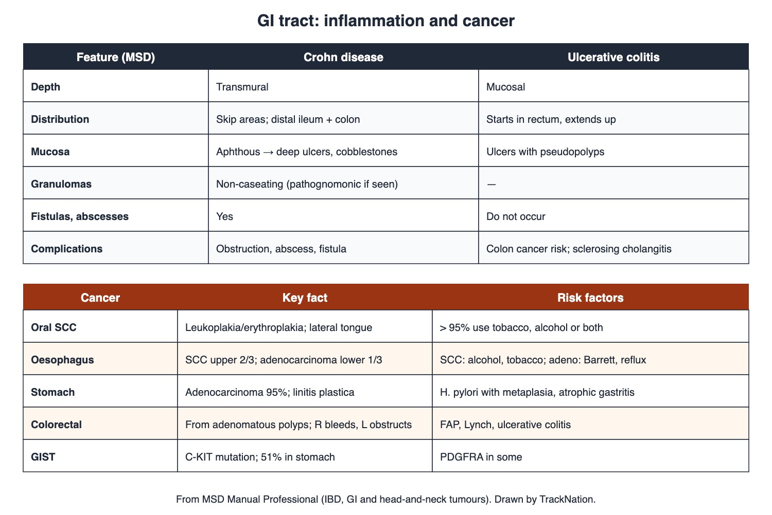
Crohn disease vs ulcerative colitis, and GI cancers. Drawn by TrackNation.
Mouth, Oesophagus and Stomach
Oral SCC: over 95% of patients use tobacco, alcohol or both. Lesions appear as leukoplakia or erythroplakia, often indurated with a rolled border; floor of mouth and lateral/ventral tongue.
Oesophageal cancer: SCC in the upper two-thirds (alcohol, tobacco; also achalasia, lye strictures, Plummer-Vinson webs); adenocarcinoma in the lower third (Barrett oesophagus from reflux, obesity). Progressive dysphagia and weight loss.
Peptic ulcer: H. pylori and NSAIDs break mucosal defence. Duodenal ulcer pain is absent on waking, appears mid-morning, is relieved by food and returns 2–3 hours after meals. Rarely gastrinoma (Zollinger-Ellison).
Stomach cancer: adenocarcinoma is 95%. Risk: H. pylori with intestinal metaplasia, autoimmune atrophic gastritis, smoking, polyps. Linitis plastica: diffuse infiltration with fibrosis — a rigid "leather bottle" stomach. Symptoms appear late.
Intestine
Crohn disease: transmural, any part of the gut (usually distal ileum and colon) with skip areas; aphthous ulcers become deep ulcers with a cobblestone pattern; non-caseating granulomas are pathognomonic when seen but often absent; abscesses, fistulas, obstruction.
Ulcerative colitis: mucosal, starts in the rectum and extends up; bloody diarrhoea; pseudopolyps; no fistulas or abscesses; raised colon cancer risk; raised ALP and GGT suggest primary sclerosing cholangitis; megacolon = transverse colon > 6 cm.
Coeliac disease: gluten intolerance → villous atrophy and malabsorption. tTG-IgA and endomysial IgA are > 90% sensitive and specific; absence of HLA-DQ2/DQ8 effectively rules it out. Tropical sprue (southern India, Caribbean, South-East Asia): malabsorption with megaloblastic anaemia; tetracycline plus folate for 6 months. Whipple disease: Tropheryma whipplei — arthritis, weight loss, abdominal pain, diarrhoea.
Appendicitis: luminal obstruction (usually lymphoid hyperplasia; sometimes faecolith, foreign body, tumour, worms) → distension, bacterial overgrowth, ischaemia; untreated → gangrene and perforation.
Colorectal cancer: arises in adenomatous polyps; about 20% have an inherited component (FAP, Lynch, Peutz-Jeghers, juvenile and MUTYH polyposis); ulcerative colitis raises risk. Right-sided tumours bleed (anaemia); left-sided obstruct. CEA is raised in only about a third. GIST: C-KIT (sometimes PDGFRA) mutation; about half in the stomach.
Traps the Examiner Sets
Upper oesophagus = SCC; lower = adenocarcinoma (Barrett).
Crohn = transmural, skip, granulomas, fistulas; UC = mucosal, continuous from rectum, pseudopolyps.
Right colon cancer → anaemia; left → obstruction.
Linitis plastica = leather-bottle stomach.
Duodenal ulcer pain is relieved by food.
Memory Hooks & Shortcuts
Shortcuts to lock in this article's numbers and lists. Each one only re-packages facts proven above.
🧠 Squamous on top, Adeno at the bottom — oesophageal cancer by site
🧠 Crohn's: Cobblestones, Granulomas, Skips, Fistulas — goes through all layers — Crohn features
🧠 UC: Up from the rectum, Continuous, mucosa only — UC features
🧠 Right Red (bleeds), Left Locks (obstructs) — colon cancer sides
🧠 Duodenal Delights in Dinner (food relieves) — duodenal ulcer pain
🧠 KIT keeps GIST — GIST mutation
Rapid-Revision Cards
Oral SCC
> 95% tobacco/alcohol; leukoplakia, erythroplakia; lateral tongue.
Oesophageal cancer by site
Upper 2/3 SCC; lower 1/3 adenocarcinoma.
SCC vs adeno risks
Alcohol, tobacco vs Barrett, reflux, obesity.
Peptic ulcer causes
H. pylori, NSAIDs; rarely gastrinoma.
Duodenal ulcer pain
Mid-morning, relieved by food, back 2–3 h later.
Gastric cancer
Adenocarcinoma 95%; H. pylori + metaplasia; atrophic gastritis.
Linitis plastica
Leather-bottle stomach.
Crohn
Transmural, skip areas, cobblestones, granulomas, fistulas.
UC
Mucosal, from rectum, pseudopolyps, no fistulas.
UC + high ALP/GGT
Primary sclerosing cholangitis.
Megacolon
Transverse colon > 6 cm.
Coeliac tests
tTG-IgA, EMA > 90%; no DQ2/DQ8 rules out.
Tropical sprue
S India; megaloblastic anaemia; tetracycline + folate.
Whipple
Tropheryma whipplei; arthritis, weight loss.
Appendicitis
Lymphoid hyperplasia blocks lumen.
Colon cancer sides
Right bleeds; left obstructs.
CRC origin
Adenomatous polyps; 20% inherited.
CEA in colon cancer
~34% raised; not for screening.
GIST
C-KIT (or PDGFRA); stomach ~51%.
Sources
Facts in this article were checked against these documents.
MSD Manual Professional: Oral Squamous Cell Carcinoma; Esophageal Cancer; Stomach Cancer; Colorectal Cancer; GISTs — Merck & Co. (MSD Manuals), reviewed 2025–26 (link)
MSD Manual Professional: Peptic Ulcer Disease; Crohn Disease; Ulcerative Colitis; Celiac Disease; Tropical Sprue; Whipple Disease; Appendicitis — Merck & Co. (MSD Manuals), reviewed 2025–26 (link)
Competency Based Undergraduate Curriculum, Pathology (PA 23) — National Medical Commission, 2024
Test Yourself: Practice Questions
Thirty questions in NEET PG style, most of them clinical vignettes.
Practice Questions (NEET PG pattern)
Original questions written in the style of the exam: +4 for a correct answer, −1 for a wrong one. Every answer was checked against the sources listed above. NEET PG papers are not published by NBEMS, so these are not past-paper questions.
Q1. A man who smokes and drinks heavily has a firm ulcerated lesion with a rolled border on the lateral tongue. The likely diagnosis is
(a) aphthous ulcer
(b) oral squamous cell carcinoma
(c) lichen planus
(d) oral candidiasis
Show answer
Answer: (b) — Over 95% of patients use tobacco, alcohol or both.
Q2. Leukoplakia and erythroplakia in the mouth are recognised presentations of
(a) oral squamous cell carcinoma (leukoplakia, erythroplakia)
(b) vitamin C deficiency
(c) measles
(d) herpes simplex
Show answer
Answer: (a) — Early lesions are often painless — hence screening.
Q3. A smoker and drinker has progressive dysphagia; biopsy of a mid-oesophageal mass is most likely
(a) squamous cell carcinoma
(b) GIST
(c) lymphoma
(d) adenocarcinoma
Show answer
Answer: (a) — SCC is commonest in the upper two-thirds.
Q4. A man with years of reflux and Barrett oesophagus develops a cancer at the gastro-oesophageal junction. It is most likely
(a) leiomyoma
(b) adenocarcinoma
(c) squamous cell carcinoma
(d) small cell carcinoma
Show answer
Answer: (b) — Adenocarcinoma dominates the distal third.
Q5. Which is a recognised risk factor for oesophageal cancer listed by MSD?
(a) hepatitis B
(b) lye ingestion with stricture
(c) H. pylori eradication
(d) vitamin D excess
Show answer
Answer: (b) — Also achalasia, HPV and Plummer-Vinson webs.
Q6. The two main factors that disrupt gastroduodenal mucosal defence in peptic ulcer disease are
(a) stress and spicy food
(b) H. pylori and NSAIDs
(c) alcohol and coffee
(d) smoking and milk
Show answer
Answer: (b) — Zollinger-Ellison gastrinoma is rare.
Q7. Epigastric pain that is absent on waking, starts mid-morning, is relieved by food and returns 2–3 hours after meals suggests
(a) duodenal ulcer
(b) pancreatitis
(c) biliary colic
(d) gastric cancer
Show answer
Answer: (a) — Night pain is also common in duodenal ulcer.
Q8. A patient has multiple refractory peptic ulcers from very high gastrin levels. The cause is
(a) pernicious anaemia
(b) H. pylori only
(c) NSAID use
(d) gastrinoma (Zollinger-Ellison syndrome)
Show answer
Answer: (d) — Very few ulcer patients have it.
Q9. The commonest malignant tumour of the stomach is
(a) leiomyosarcoma
(b) adenocarcinoma (95%)
(c) lymphoma
(d) GIST
Show answer
Answer: (b) — MSD.
Q10. Endoscopy shows a rigid, thick-walled stomach; biopsy shows tumour infiltrating the wall with fibrosis. This is
(a) gastric ulcer
(b) Ménétrier disease
(c) linitis plastica (leather-bottle stomach)
(d) GIST
Show answer
Answer: (c) — A diffusely infiltrating gastric cancer.
Q11. A recognised risk factor for gastric adenocarcinoma is
(a) hepatitis C
(b) H. pylori with extensive intestinal metaplasia
(c) coeliac disease
(d) duodenal ulcer
Show answer
Answer: (b) — Also autoimmune atrophic gastritis and smoking.
Q12. A patient has diarrhoea, abdominal pain and an enteric fistula; diseased bowel segments are sharply separated from normal bowel. The likely diagnosis is
(a) irritable bowel syndrome
(b) ulcerative colitis
(c) amoebic colitis
(d) Crohn disease
Show answer
Answer: (d) — Transmural inflammation with skip areas.
Q13. Biopsy of inflamed ileum shows non-caseating granulomas in the bowel wall. This favours
(a) Crohn disease
(b) ulcerative colitis
(c) coeliac disease
(d) tuberculosis always
Show answer
Answer: (a) — Pathognomonic when present, but often absent.
Q14. Deep longitudinal and transverse ulcers separated by swollen mucosa give the Crohn bowel a
(a) lead-pipe appearance
(b) cobblestone appearance
(c) apple-core appearance
(d) string-of-pearls appearance
Show answer
Answer: (b) — MSD.
Q15. A patient has bloody diarrhoea; colonoscopy shows continuous mucosal inflammation starting at the rectum. The likely diagnosis is
(a) ischaemic colitis
(b) diverticulitis
(c) ulcerative colitis
(d) Crohn disease
Show answer
Answer: (c) — UC is mucosal and starts in the rectum.
Q16. Islands of regenerating mucosa projecting above ulcerated areas in ulcerative colitis are called
(a) adenomas
(b) pseudopolyps
(c) granulomas
(d) hamartomas
Show answer
Answer: (b) — Fistulas and abscesses do not occur in UC.
Q17. Which feature points to Crohn disease rather than ulcerative colitis?
(a) fistulas and abscesses
(b) rectal involvement
(c) bloody diarrhoea
(d) pseudopolyps
Show answer
Answer: (a) — MSD: fistulas and abscesses do not occur in UC.
Q18. A patient with ulcerative colitis has raised alkaline phosphatase and GGT. Suspect
(a) Gilbert syndrome
(b) primary sclerosing cholangitis
(c) gallstones only
(d) hepatitis A
Show answer
Answer: (b) — UC also raises long-term colon cancer risk.
Q19. A woman with diarrhoea and iron deficiency has villous atrophy on duodenal biopsy. The most useful blood test is
(a) CEA
(b) stool culture
(c) ANA
(d) tissue transglutaminase IgA (and endomysial IgA)
Show answer
Answer: (d) — Both > 90% sensitive and specific.
Q20. In suspected coeliac disease with conflicting biopsy and serology, a negative test for HLA-DQ2 and DQ8
(a) means lymphoma
(b) effectively rules coeliac disease out
(c) is irrelevant
(d) confirms it
Show answer
Answer: (b) — High sensitivity, low specificity.
Q21. A man from southern India has chronic diarrhoea with malabsorption and megaloblastic anaemia. Coeliac serology is negative. Likely diagnosis and treatment:
(a) tropical sprue — tetracycline plus folate for 6 months
(b) coeliac disease — gluten-free diet
(c) Whipple disease — no treatment
(d) giardiasis — metronidazole once
Show answer
Answer: (a) — Endemic in southern India, Caribbean, South-East Asia.
Q22. A middle-aged man has arthritis, weight loss, diarrhoea and abdominal pain; small-bowel biopsy shows the organism. The bacterium is
(a) Clostridioides difficile
(b) Tropheryma whipplei
(c) Giardia
(d) Mycobacterium avium
Show answer
Answer: (b) — Whipple disease.
Q23. The usual cause of luminal obstruction in acute appendicitis is
(a) lymphoid hyperplasia
(b) worms
(c) foreign body
(d) carcinoid tumour
Show answer
Answer: (a) — Occasionally faecolith, foreign body, tumour or worms.
Q24. An elderly woman has unexplained iron-deficiency anaemia and occult blood in stool; colonoscopy finds a caecal mass. Right-sided colon cancers typically present with
(a) jaundice
(b) bleeding and anaemia
(c) early obstruction
(d) pencil-thin stools
Show answer
Answer: (b) — Left-sided tumours tend to obstruct.
Q25. Most colorectal cancers arise from
(a) normal mucosa directly in all cases
(b) adenomatous polyps
(c) juvenile polyps
(d) hyperplastic polyps
Show answer
Answer: (b) — About 80% sporadic, 20% inherited component.
Q26. Which hereditary syndrome predisposes to colorectal cancer?
(a) Turner syndrome
(b) Gilbert syndrome
(c) Marfan syndrome
(d) Lynch syndrome
Show answer
Answer: (d) — Also FAP, Peutz-Jeghers, juvenile and MUTYH polyposis.
Q27. In colon cancer, a raised serum CEA is found in about
(a) 90% of early cancers
(b) no patients
(c) a third of patients — so it is not a screening test
(d) all patients
Show answer
Answer: (c) — 34% in a US stage I–III study.
Q28. A submucosal gastric tumour shows a mutation in C-KIT. The tumour is a
(a) gastrointestinal stromal tumour (GIST)
(b) lymphoma
(c) adenocarcinoma
(d) carcinoid
Show answer
Answer: (a) — Some have PDGFRA mutations; about half arise in the stomach.
Q29. The commonest site of GIST is the
(a) appendix
(b) oesophagus
(c) stomach
(d) rectum
Show answer
Answer: (c) — 51% stomach, 36% small bowel in one study.
Q30. In severe ulcerative colitis, megacolon is defined radiologically as a transverse colon diameter above
(a) 10 cm
(b) 15 cm
(c) 3 cm
(d) 6 cm
Show answer
Answer: (d) — Barium enema is avoided in severe flares (perforation risk).
Active Recall
From memory: give the risk factors of oral cancer; compare oesophageal SCC and adenocarcinoma; list peptic ulcer causes and duodenal pain pattern; describe gastric cancer and linitis plastica; contrast Crohn disease and ulcerative colitis; give coeliac serology, tropical sprue treatment and the cause of appendicitis; and describe colorectal cancer by side, its origins and GIST.