Haematology Basics and Microcytic Anaemia for NEET PG Pathology: Iron Deficiency, Anaemia of Chronic Disease, Thalassaemia Trait
Start Here
🎯 Exam priority: Very high. Expect: a woman with fatigue and low ferritin, a microcytic picture with high RBC count (thalassaemia trait), anaemia of chronic disease in rheumatoid arthritis, ring sideroblasts, and the tube colour for a blood count.
CBME mapping: PA 13.1–13.4, 14.1–14.2. Indian cut-offs: Anemia Mukt Bharat.
Making Red Cells
Red cells are made in the marrow under erythropoietin, produced by peritubular interstitial fibroblasts of the kidney in response to low oxygen delivery.
They live about 120 days; reticulocytes make up 0.5–2.5% of adult red cells.
Extramedullary haematopoiesis (mainly in the spleen) is seen in myelofibrosis, which also gives nucleated and teardrop red cells.
Anaemia: Definition and Work-up
Anaemia is a fall in red-cell number, lowering haematocrit and haemoglobin. Start with a CBC, reticulocyte count and peripheral smear.
Microcytic if MCV < 80 fL; macrocytic if MCV > 100 fL (many reticulocytes can raise the MCV). The reticulocyte count shows how well the marrow is compensating.
Tubes (WHO): EDTA, purple top — haematology and cross-match; citrate, light blue — coagulation tests. Both work by binding calcium.
Indian cut-offs for anaemia (Hb, g/dL; Anemia Mukt Bharat):
Group | Mild | Moderate | Severe |
Children 6–59 months | 10–10.9 | 7–9.9 | < 7 |
Pregnant women | 10–10.9 | 7–9.9 | < 7 |
Non-pregnant women (≥ 15 y) | 11–11.9 | 8–10.9 | < 8 |
Men (≥ 15 y) | 11–12.9 | 8–10.9 | < 8 |
Iron Deficiency
The commonest cause of anaemia, usually from blood loss. Iron is absorbed in the duodenum and upper jejunum, best as haem iron; low stores suppress hepcidin, raising absorption.
Stages: stores fall first (ferritin < 30 ng/mL with normal Hb), then iron-starved red-cell production, then microcytosis and hypochromia.
Labs: low ferritin and serum iron, high TIBC. Smear: anisocytosis, poikilocytosis; cigar-shaped cells in severe cases; no polychromasia or basophilic stippling.
Clues: pica; spoon-shaped nails (koilonychia). In men and postmenopausal women without obvious loss, examine the GI tract.
Response to iron: reticulocytes rise after about 1 week; Hb starts rising after 1–2 weeks.
Telling Microcytic Anaemias Apart
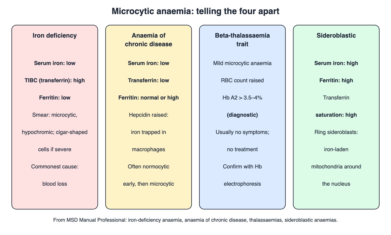
Iron deficiency, anaemia of chronic disease, beta-thalassaemia trait and sideroblastic anaemia compared. Drawn by TrackNation.
Anaemia of chronic disease: raised hepcidin traps iron in macrophages; serum iron and transferrin low, ferritin normal or high; normocytic early, then microcytic.
Beta-thalassaemia trait: mild microcytic anaemia with a raised red-cell count; Hb A2 > 3.5–4% is diagnostic (normal ≤ 2.5%); no treatment needed.
Sideroblastic anaemia: iron cannot enter protoporphyrin IX; high serum iron, ferritin and saturation; ring sideroblasts. Acquired forms are usually part of myelodysplastic syndrome.
Traps the Examiner Sets
Low ferritin = iron deficiency; normal/high ferritin with low iron = chronic disease.
Microcytosis with a high RBC count suggests thalassaemia trait, not iron deficiency.
Iron deficiency in an older man: look for a GI cancer.
Purple (EDTA) for counts, light blue (citrate) for clotting.
Memory Hooks & Shortcuts
Shortcuts to lock in this article's numbers and lists. Each one only re-packages facts proven above.
🧠 Ferritin Falls First — the earliest sign of iron deficiency
🧠 FIT test: Ferritin low, Iron low, TIBC high — iron deficiency labs
🧠 Chronic disease Clings to iron (hepcidin) — ferritin normal or high
🧠 Thal trait: Tiny cells, Too many (high RBC), A2 Above 3.5 — beta-thalassaemia minor
🧠 Ring around the nucleus = Sideroblastic — ring sideroblasts
🧠 Purple for counting, Blue for clotting — EDTA vs citrate tubes
Rapid-Revision Cards
Erythropoietin source
Kidney peritubular interstitial fibroblasts.
RBC lifespan
~120 days; reticulocytes 0.5–2.5%.
Extramedullary haematopoiesis
Spleen; myelofibrosis (teardrop cells).
Anaemia work-up
CBC, reticulocyte count, smear.
MCV cut-offs
Microcytic < 80 fL; macrocytic > 100 fL.
EDTA tube
Purple; CBC, cross-match.
Citrate tube
Light blue; coagulation.
AMB cut-offs (pregnancy)
Mild 10–10.9, moderate 7–9.9, severe < 7.
AMB cut-offs (men)
Mild 11–12.9, moderate 8–10.9, severe < 8.
Commonest anaemia
Iron deficiency (blood loss).
Iron absorption
Duodenum, upper jejunum; haem best; hepcidin falls.
Earliest iron change
Ferritin < 30 with normal Hb.
IDA labs
Low ferritin, low iron, high TIBC.
IDA smear
Aniso-poikilocytosis; cigar cells; no stippling.
IDA clues
Pica, koilonychia.
IDA in older man
Look for GI cancer.
Iron response
Retics at ~1 wk; Hb rises 1–2 wk.
ACD
High hepcidin; low iron and transferrin; ferritin normal/high.
Beta-thal trait
Microcytic, high RBC count; Hb A2 > 3.5–4%.
Sideroblastic
High iron/ferritin; ring sideroblasts; MDS.
Sources
Facts in this article were checked against these documents.
MSD Manual Professional: Red Blood Cell Production; Etiology and Evaluation of Anemia; Iron Deficiency Anemia; Anemia of Chronic Disease; Sideroblastic Anemias; Thalassemias; Primary Myelofibrosis — Merck & Co. (MSD Manuals), reviewed 2025–2026 (link)
WHO guidelines on drawing blood: best practices in phlebotomy — World Health Organization, 2010 (link)
Anemia Mukt Bharat: Operational Guidelines — Ministry of Health and Family Welfare, 2018
Competency Based Undergraduate Curriculum, Pathology (PA 13–14) — National Medical Commission, 2024
Test Yourself: Practice Questions
Twenty-four questions in NEET PG style, most of them clinical vignettes.
Practice Questions (NEET PG pattern)
Original questions written in the style of the exam: +4 for a correct answer, −1 for a wrong one. Every answer was checked against the sources listed above. NEET PG papers are not published by NBEMS, so these are not past-paper questions.
Q1. A 32-year-old woman with heavy periods has Hb 9 g/dL, MCV 70 fL, low serum ferritin and high TIBC. The diagnosis is
(a) beta-thalassaemia trait
(b) sideroblastic anaemia
(c) anaemia of chronic disease
(d) iron deficiency anaemia
Show answer
Answer: (d) — Low ferritin and serum iron with high TIBC.
Q2. The commonest cause of anaemia, usually due to blood loss, is
(a) iron deficiency
(b) thalassaemia
(c) vitamin B12 deficiency
(d) aplastic anaemia
Show answer
Answer: (a) — MSD.
Q3. The earliest laboratory change as iron stores fall, while haemoglobin is still normal, is
(a) microcytosis
(b) a fall in serum ferritin (< 30 ng/mL)
(c) a fall in Hb
(d) hypochromia
Show answer
Answer: (b) — Stores deplete first; microcytosis and hypochromia come in stage 3.
Q4. Iron is absorbed mainly in the
(a) terminal ileum
(b) stomach
(c) colon
(d) duodenum and upper jejunum
Show answer
Answer: (d) — Haem iron (meat) is absorbed best.
Q5. A woman with a long-standing autoimmune disease has mild anaemia, low serum iron, low transferrin and a high ferritin. The mechanism is
(a) B12 deficiency
(b) blood loss
(c) ineffective globin synthesis
(d) raised hepcidin sequestering iron (anaemia of chronic disease)
Show answer
Answer: (d) — Hepcidin blocks absorption and recycling; ferritin normal or high.
Q6. A 25-year-old has Hb 11.5 g/dL, MCV 64 fL and a raised red-cell count; he feels well. The likely diagnosis is
(a) severe iron deficiency
(b) aplastic anaemia
(c) anaemia of chronic disease
(d) beta-thalassaemia trait
Show answer
Answer: (d) — Trait: microcytic anaemia with elevated RBC count, usually asymptomatic.
Q7. In that man, the finding diagnostic of beta-thalassaemia minor is
(a) Hb F of 90%
(b) Hb A2 above 3.5–4%
(c) ring sideroblasts
(d) low ferritin
Show answer
Answer: (b) — Normal Hb A2 ≤ 2.5%.
Q8. Beta-thalassaemia trait usually needs
(a) no treatment
(b) iron therapy
(c) splenectomy
(d) regular transfusion
Show answer
Answer: (a) — MSD: no intervention is needed.
Q9. A marrow iron stain shows erythroblasts with a ring of iron-laden mitochondria around the nucleus; serum iron and ferritin are high. This is
(a) anaemia of chronic disease
(b) thalassaemia trait
(c) sideroblastic anaemia
(d) iron deficiency
Show answer
Answer: (c) — Iron cannot be incorporated into protoporphyrin IX.
Q10. Most acquired sideroblastic anaemias are part of
(a) hypothyroidism
(b) thalassaemia
(c) iron deficiency
(d) myelodysplastic syndrome
Show answer
Answer: (d) — MSD.
Q11. A 65-year-old man with no obvious bleeding has iron deficiency anaemia. The next step is
(a) evaluate the GI tract for occult cancer
(b) oral iron only and no tests
(c) bone marrow biopsy
(d) Hb electrophoresis
Show answer
Answer: (a) — Anaemia may be the only sign of occult GI cancer.
Q12. A child with iron deficiency craves and eats non-food substances. This is called
(a) glossitis
(b) koilonychia
(c) pica
(d) dysphagia
Show answer
Answer: (c) — MSD: suspect iron deficiency, particularly if pica is present.
Q13. Spoon-shaped, concave nails in an anaemic woman suggest
(a) haemolysis
(b) B12 deficiency
(c) thalassaemia
(d) iron deficiency (koilonychia)
Show answer
Answer: (d) — MSD image caption.
Q14. In severe iron deficiency, the smear characteristically shows
(a) anisocytosis, poikilocytosis and cigar-shaped cells, without polychromasia
(b) target cells with basophilic stippling
(c) macro-ovalocytes
(d) spherocytes
Show answer
Answer: (a) — Polychromasia and basophilic stippling are conspicuously absent.
Q15. After starting oral iron, the first sign of response is
(a) Hb normal at 3 days
(b) MCV normal at 1 week
(c) ferritin normal at 1 day
(d) a reticulocyte rise after about 1 week
Show answer
Answer: (d) — Hb begins to rise after 1–2 weeks.
Q16. A red-cell population is called microcytic when MCV is below
(a) 60 fL
(b) 100 fL
(c) 80 fL
(d) 90 fL
Show answer
Answer: (c) — Macrocytic is > 100 fL.
Q17. A patient bleeding acutely has a high reticulocyte count and a slightly raised MCV. The MCV is raised because
(a) reticulocytes are larger than mature red cells
(b) of lab error
(c) of iron deficiency
(d) of B12 deficiency
Show answer
Answer: (a) — Large numbers of reticulocytes can elevate the MCV.
Q18. A blood sample for a complete blood count should be collected in the tube with
(a) EDTA (purple top)
(b) clot activator (red top)
(c) fluoride (grey top)
(d) citrate (light blue top)
Show answer
Answer: (a) — WHO: EDTA for haematology and cross-match; citrate for coagulation.
Q19. For a prothrombin time, blood is collected in
(a) EDTA (purple top)
(b) a plain tube
(c) heparin
(d) sodium citrate (light blue top)
Show answer
Answer: (d) — WHO phlebotomy guideline.
Q20. Erythropoietin is produced mainly by
(a) the spleen
(b) hepatocytes
(c) peritubular interstitial fibroblasts of the kidney
(d) bone marrow stromal cells
Show answer
Answer: (c) — In response to decreased oxygen delivery.
Q21. A man with splenomegaly and marrow fibrosis has teardrop and nucleated red cells on his smear. The spleen is enlarged partly from
(a) iron overload
(b) thalassaemia trait
(c) B12 deficiency
(d) extramedullary haematopoiesis (myelofibrosis)
Show answer
Answer: (d) — MSD: primary myelofibrosis.
Q22. Under Anemia Mukt Bharat, a pregnant woman with Hb 6.5 g/dL has
(a) mild anaemia
(b) severe anaemia (< 7 g/dL)
(c) moderate anaemia
(d) no anaemia
Show answer
Answer: (b) — Pregnant women: mild 10–10.9, moderate 7–9.9, severe < 7.
Q23. Under Anemia Mukt Bharat, a man with Hb 12 g/dL has
(a) mild anaemia (11–12.9)
(b) no anaemia
(c) severe anaemia
(d) moderate anaemia
Show answer
Answer: (a) — Men ≥ 15 y: mild 11–12.9, moderate 8–10.9, severe < 8.
Q24. Normal adult red cells survive about
(a) 30 days
(b) 1 year
(c) 7 days
(d) 120 days
Show answer
Answer: (d) — Then cleared mainly by the spleen.
Active Recall
From memory: explain red-cell production and lifespan, the first tests for anaemia and the MCV cut-offs, the sample tubes, the stages and lab picture of iron deficiency, and how anaemia of chronic disease, beta-thalassaemia trait and sideroblastic anaemia differ from it.