Pleura, Pulmonary Embolism and Interstitial Lung Disease for NEET PG Medicine: Light's Criteria, Pneumothorax, PE, IPF, Sarcoidosis
Start Here
🎯 Exam priority: Medium–high. Expect: Light's criteria numbers, causes of transudates vs exudates, empyema drainage triggers, tension pneumothorax first step, PE investigations and ECG/X-ray signs, IPF crackles and treatment, Löfgren vs Heerfordt, and the pulmonary hypertension cut-off.
CBME mapping: respiratory competencies in GM 3 and GM 28. Tuberculous effusion is in article 7; PE pathology (Virchow triad) in Pathology article 3.
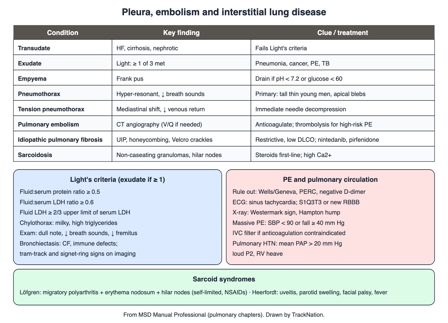
Pleural effusion, pneumothorax, PE, IPF, sarcoidosis. Drawn by TrackNation.
Pleural Effusion
Signs: reduced breath sounds, dullness to percussion, absent tactile fremitus on the affected side.
Transudates: heart failure, cirrhosis with ascites, nephrotic syndrome. Exudates: pneumonia, cancer, pulmonary embolism, tuberculosis.
Chylothorax: milky fluid high in triglycerides (trauma, surgery, tumours). Empyema: frank pus; drain completely if pH < 7.20, glucose < 60 mg/dL, positive Gram stain or culture, or loculations.
Light's criteria — exudate if ≥ 1 | Cut-off |
Pleural fluid : serum protein | ≥ 0.5 |
Pleural fluid : serum LDH | ≥ 0.6 |
Pleural fluid LDH | ≥ 2/3 of the upper limit of serum LDH |
Pneumothorax
Primary spontaneous: no lung disease; classically tall, thin men in their teens and 20s; rupture of apical blebs (smoking). Signs: absent fremitus, hyper-resonance, reduced breath sounds.
Tension pneumothorax: positive intrapleural pressure throughout breathing collapses the lung, shifts the mediastinum and impairs venous return → shock. Immediate needle decompression.
Asymptomatic primary spontaneous pneumothorax: observe with a repeat X-ray regardless of size; symptomatic: aspiration or drain devices.
Pulmonary Embolism and Pulmonary Hypertension
Work-up: clinical scores (Wells, revised Geneva, PERC); a negative D-dimer makes PE very unlikely in low-probability patients; CT angiography is the usual test (V/Q scan sometimes).
Signs: ECG often sinus tachycardia; S1Q3T3 or new RBBB suggests RV strain. X-ray classics: Westermark sign (focal oligaemia) and Hampton hump (peripheral wedge).
Treatment: anticoagulation; thrombolysis (some reserve it for high-risk PE) or catheter/surgical clot removal; IVC filter if anticoagulation is contraindicated.
Pulmonary hypertension: mean pulmonary arterial pressure > 20 mm Hg; signs include a loud P2 and RV heave.
PE risk class (MSD) | Definition |
High risk (massive) | RV dysfunction with hypotension: SBP < 90 or fall ≥ 40 mm Hg for 15 minutes |
Intermediate (submassive) | RV dysfunction without hypotension (imaging, biomarkers) |
Low risk | No RV dysfunction, normotensive |
Interstitial Lung Disease, Sarcoidosis, Bronchiectasis
Idiopathic pulmonary fibrosis = usual interstitial pneumonia: exertional breathlessness, cough, fine bibasal Velcro crackles, clubbing in about half; honeycombing; restrictive spirometry with low diffusing capacity. Treat with pirfenidone or nintedanib, oxygen, rehabilitation, transplant.
Sarcoidosis: non-caseating granulomas, mostly lungs and thoracic nodes (hilar/mediastinal in 90%); uveitis is the commonest eye lesion; hypercalcaemia from macrophage activation of vitamin D. A normal chest X-ray (stage 0) does not exclude it. First-line treatment: corticosteroids. Löfgren syndrome (arthritis, erythema nodosum, hilar nodes) is self-limited — NSAIDs. Heerfordt syndrome (uveoparotid fever): uveitis, parotid swelling, facial palsy, fever.
Bronchiectasis: cystic fibrosis, immune defects, recurrent infection; imaging shows tram-track lines and the signet-ring sign.
Traps the Examiner Sets
Effusion = dull; pneumothorax = hyper-resonant.
Tension pneumothorax: decompress before the X-ray.
Light's: 0.5 protein, 0.6 LDH, 2/3 LDH — any one = exudate.
S1Q3T3 is suggestive, not common; sinus tachycardia is commoner.
Löfgren resolves on its own; Heerfordt = uveoparotid fever.
Memory Hooks & Shortcuts
Shortcuts to lock in this article's numbers and lists. Each one only re-packages facts proven above.
🧠 Light's: 'Point five, point six, two-thirds' — Light's criteria
🧠 Dull = Drink (fluid); Drum = air (hyper-resonant) — effusion vs pneumothorax
🧠 Tension: Treat before the Test (needle first) — tension pneumothorax
🧠 Löfgren = 'LEgs, Joints, Hilum' — Leaves by itself — Löfgren
🧠 Heerfordt = 'Eyes, Ears (parotid), Face, Fever' — Heerfordt
🧠 Velcro crackles, Velcro lungs (IPF) — IPF
Rapid-Revision Cards
Transudate causes
Heart failure, cirrhosis, nephrotic syndrome.
Exudate causes
Pneumonia, cancer, PE, TB.
Light's criteria
Protein ratio ≥ 0.5; LDH ratio ≥ 0.6; LDH ≥ 2/3 ULN.
Chylothorax
Milky; high triglycerides.
Empyema drain
pH < 7.2, glucose < 60, Gram+, loculated.
Effusion signs
Dull, ↓ breath sounds, ↓ fremitus.
Primary pneumothorax
Tall thin young men; apical blebs.
Pneumothorax signs
Hyper-resonant, ↓ breath sounds.
Tension pneumothorax
Needle decompression first.
PE test of choice
CT pulmonary angiography.
D-dimer
Negative rules out in low probability.
PE ECG
Sinus tachycardia; S1Q3T3; new RBBB.
Westermark / Hampton
Oligaemia / peripheral wedge.
Massive PE
SBP < 90 or fall ≥ 40 mm Hg.
IVC filter
If anticoagulation contraindicated.
IPF
UIP, honeycomb, Velcro crackles, clubbing 50%.
IPF drugs
Pirfenidone, nintedanib.
Sarcoidosis
Non-caseating granulomas; hilar nodes 90%.
Löfgren
Arthritis + erythema nodosum + hilar nodes; NSAIDs.
Heerfordt
Uveitis, parotids, facial palsy, fever.
Sarcoid hypercalcaemia
Macrophage 1α-hydroxylation of vitamin D.
Pulmonary HTN
Mean PAP > 20 mm Hg.
Bronchiectasis imaging
Tram tracks, signet ring.
Sources
Facts in this article were checked against these documents.
MSD Manual Professional: Pleural Effusion; Pneumothorax; Pulmonary Embolism; Idiopathic Pulmonary Fibrosis; Sarcoidosis; Pulmonary Hypertension; Bronchiectasis — Merck & Co. (MSD Manuals), reviewed 2025–26 (link)
Competency Based Undergraduate Curriculum, General Medicine (GM 3, GM 28) — National Medical Commission, 2024
Test Yourself: Practice Questions
Thirty questions in NEET PG style, most of them clinical vignettes.
Practice Questions (NEET PG pattern)
Original questions written in the style of the exam: +4 for a correct answer, −1 for a wrong one. Every answer was checked against the sources listed above. NEET PG papers are not published by NBEMS, so these are not past-paper questions.
Q1. Pleural fluid protein 4.2 g/dL with serum protein 6.5 g/dL (ratio 0.65). By Light's criteria the effusion is
(a) indeterminate
(b) a transudate
(c) an exudate
(d) chylous
Show answer
Answer: (c) — Protein ratio ≥ 0.5 is enough.
Q2. Which is NOT one of Light's criteria for an exudate?
(a) fluid:serum LDH ≥ 0.6
(b) pleural fluid glucose < 60 mg/dL
(c) fluid:serum protein ≥ 0.5
(d) fluid LDH ≥ 2/3 upper limit of serum
Show answer
Answer: (b) — Low glucose is an empyema/drainage marker.
Q3. The commonest causes of a transudative pleural effusion are
(a) heart failure, cirrhosis and nephrotic syndrome
(b) pneumonia, cancer and TB
(c) trauma and surgery
(d) pancreatitis and SLE
Show answer
Answer: (a) — Exudates: pneumonia, cancer, PE, TB.
Q4. After a thoracic duct injury, pleural fluid is milky white. The fluid will be high in
(a) glucose
(b) amylase
(c) triglycerides
(d) red cells only
Show answer
Answer: (c) — Chylothorax.
Q5. A parapneumonic effusion should be completely drained when the fluid shows
(a) pH < 7.20 or glucose < 60 mg/dL
(b) pH 7.45
(c) clear straw colour
(d) protein ratio 0.3
Show answer
Answer: (a) — Also positive Gram stain/culture or loculations.
Q6. Examination shows reduced breath sounds, dullness to percussion and absent tactile fremitus at the right base. The likely finding is
(a) pleural effusion
(b) pneumothorax
(c) emphysema
(d) consolidation with patent bronchus
Show answer
Answer: (a) — Pleural thickening can mimic it.
Q7. A tall, thin 20-year-old smoker has sudden pleuritic chest pain and breathlessness; the left chest is hyper-resonant with reduced breath sounds. The diagnosis is
(a) pleural effusion
(b) pneumonia
(c) primary spontaneous pneumothorax
(d) pulmonary embolism
Show answer
Answer: (c) — Rupture of apical blebs.
Q8. A patient becomes hypotensive; the mediastinum is shifted away from a hyper-resonant hemithorax with absent breath sounds. Immediate management is
(a) needle decompression
(b) intubation only
(c) CT chest
(d) chest X-ray first
Show answer
Answer: (a) — Tension pneumothorax impairs venous return.
Q9. Tension pneumothorax causes shock mainly by
(a) bleeding into the pleura
(b) impairing venous return to the heart
(c) sepsis
(d) direct myocardial injury
Show answer
Answer: (b) — Positive intrapleural pressure throughout the cycle.
Q10. An asymptomatic young man has a primary spontaneous pneumothorax on X-ray. Per MSD, management is
(a) immediate surgery
(b) intercostal drain in all
(c) thrombolysis
(d) observation with a follow-up radiograph regardless of size
Show answer
Answer: (d) — Symptomatic: aspiration or ambulatory devices.
Q11. The investigation most commonly used to diagnose pulmonary embolism is
(a) echocardiography
(b) D-dimer alone
(c) CT pulmonary angiography
(d) chest X-ray
Show answer
Answer: (c) — V/Q scan sometimes.
Q12. In an outpatient with low clinical probability of PE, a negative D-dimer
(a) makes PE very unlikely
(b) requires immediate thrombolysis
(c) confirms PE
(d) is meaningless
Show answer
Answer: (a) — Use with Wells/Geneva scores.
Q13. An ECG pattern suggesting pulmonary embolism is
(a) S wave in I, Q wave and inverted T in III
(b) delta wave
(c) U waves
(d) ST elevation in II, III, aVF
Show answer
Answer: (a) — Or a new RBBB.
Q14. A peripheral wedge-shaped pleural-based opacity on chest X-ray after PE is the
(a) golden S sign
(b) Kerley B line
(c) Hampton hump
(d) Westermark sign
Show answer
Answer: (c) — Westermark = focal loss of vascular markings.
Q15. A PE patient has RV dysfunction and systolic BP 82 mm Hg. The risk class is
(a) not PE
(b) high risk (massive)
(c) intermediate (submassive)
(d) low risk
Show answer
Answer: (b) — Thrombolysis is considered.
Q16. A patient with acute PE has a recent intracranial haemorrhage, so anticoagulation is contraindicated. An option is
(a) an inferior vena cava filter
(b) aspirin alone
(c) high-dose thrombolysis
(d) no treatment
Show answer
Answer: (a) — Until anticoagulation can resume.
Q17. A 68-year-old has progressive exertional breathlessness, dry cough, fine bibasal 'Velcro' crackles and clubbing; HRCT shows honeycombing. The diagnosis is
(a) bronchiectasis
(b) idiopathic pulmonary fibrosis
(c) sarcoidosis
(d) COPD
Show answer
Answer: (b) — Histology: usual interstitial pneumonia.
Q18. Antifibrotic drugs used in idiopathic pulmonary fibrosis are
(a) salbutamol and tiotropium
(b) pirfenidone and nintedanib
(c) methotrexate
(d) prednisolone and azathioprine as first line
Show answer
Answer: (b) — With oxygen, rehabilitation, transplant.
Q19. Spirometry in idiopathic pulmonary fibrosis shows
(a) a restrictive pattern with reduced diffusing capacity
(b) reversible obstruction
(c) obstruction with FEV1/FVC < 0.7
(d) normal values always
Show answer
Answer: (a) — Fibrosis stiffens the lungs.
Q20. A young woman has fever, painful ankles, red tender shin nodules and bilateral hilar lymphadenopathy. The likely diagnosis and treatment are
(a) rheumatic fever — penicillin
(b) Löfgren syndrome — usually NSAIDs; self-limited
(c) TB — start ATT
(d) lymphoma — chemotherapy
Show answer
Answer: (b) — Arthritis, erythema nodosum, hilar nodes.
Q21. A patient has uveitis, bilateral parotid swelling, facial nerve palsy and fever. This is
(a) mumps
(b) Heerfordt syndrome (uveoparotid fever) of sarcoidosis
(c) Sjögren syndrome
(d) Bell palsy only
Show answer
Answer: (b) — A sarcoid variant.
Q22. Hypercalcaemia in sarcoidosis results from
(a) increased activation of vitamin D by granuloma macrophages
(b) parathyroid adenoma
(c) bone metastases
(d) milk-alkali syndrome
Show answer
Answer: (a) — 1,25-dihydroxyvitamin D production.
Q23. The histological hallmark of sarcoidosis is
(a) Reed-Sternberg cells
(b) non-caseating granulomas
(c) Schaumann bodies only
(d) caseating granulomas
Show answer
Answer: (b) — Lungs and lymphatic system most often affected.
Q24. First-line treatment for symptomatic pulmonary sarcoidosis is
(a) corticosteroids
(b) antitubercular drugs
(c) antifibrotics
(d) antibiotics
Show answer
Answer: (a) — Löfgren syndrome usually needs only NSAIDs.
Q25. The commonest ocular manifestation of sarcoidosis is
(a) retinal detachment
(b) uveitis
(c) cataract
(d) glaucoma
Show answer
Answer: (b) — Blurred vision, photophobia, tearing.
Q26. A normal chest X-ray in suspected cardiac sarcoidosis
(a) confirms TB
(b) does not exclude sarcoidosis
(c) means stage 4
(d) excludes sarcoidosis
Show answer
Answer: (b) — Stage 0; HRCT is more sensitive.
Q27. Pulmonary hypertension is defined haemodynamically by a mean pulmonary arterial pressure above
(a) 40 mm Hg
(b) 20 mm Hg
(c) 60 mm Hg
(d) 10 mm Hg
Show answer
Answer: (b) — Wedge ≤ 15 mm Hg in pre-capillary disease.
Q28. A thickened, dilated airway seen next to its adjacent artery on CT is the
(a) halo sign
(b) signet-ring sign of bronchiectasis
(c) tree-in-bud
(d) crazy paving
Show answer
Answer: (b) — Tram-track lines are the other classic sign.
Q29. Bronchiectasis is commonly caused by all EXCEPT
(a) sarcoid hypercalcaemia
(b) immune defects
(c) cystic fibrosis
(d) recurrent infections
Show answer
Answer: (a) — Some cases are idiopathic.
Q30. Clubbing in idiopathic pulmonary fibrosis is present in about
(a) 50% of cases
(b) never
(c) all cases
(d) 5%
Show answer
Answer: (a) — Velcro crackles are the classic sign.
Active Recall
From memory: list causes of transudates and exudates and Light's criteria; describe chylothorax and empyema drainage; describe primary and tension pneumothorax and their management; outline PE work-up, signs, risk classes and treatment; and describe IPF, sarcoidosis syndromes and the pulmonary hypertension cut-off.